
Integumentary System
Your skin is the largest organ in your body, weighing about 4 kg (roughly 9 pounds) and covering approximately 2 square meters. It is far more than a passive wrapper - it is an active barrier, sensory organ, immune defense, vitamin D factory, and thermoregulation system all rolled into one.
The Three Layers of Skin
| Layer | Type of Tissue | Key Features |
|---|---|---|
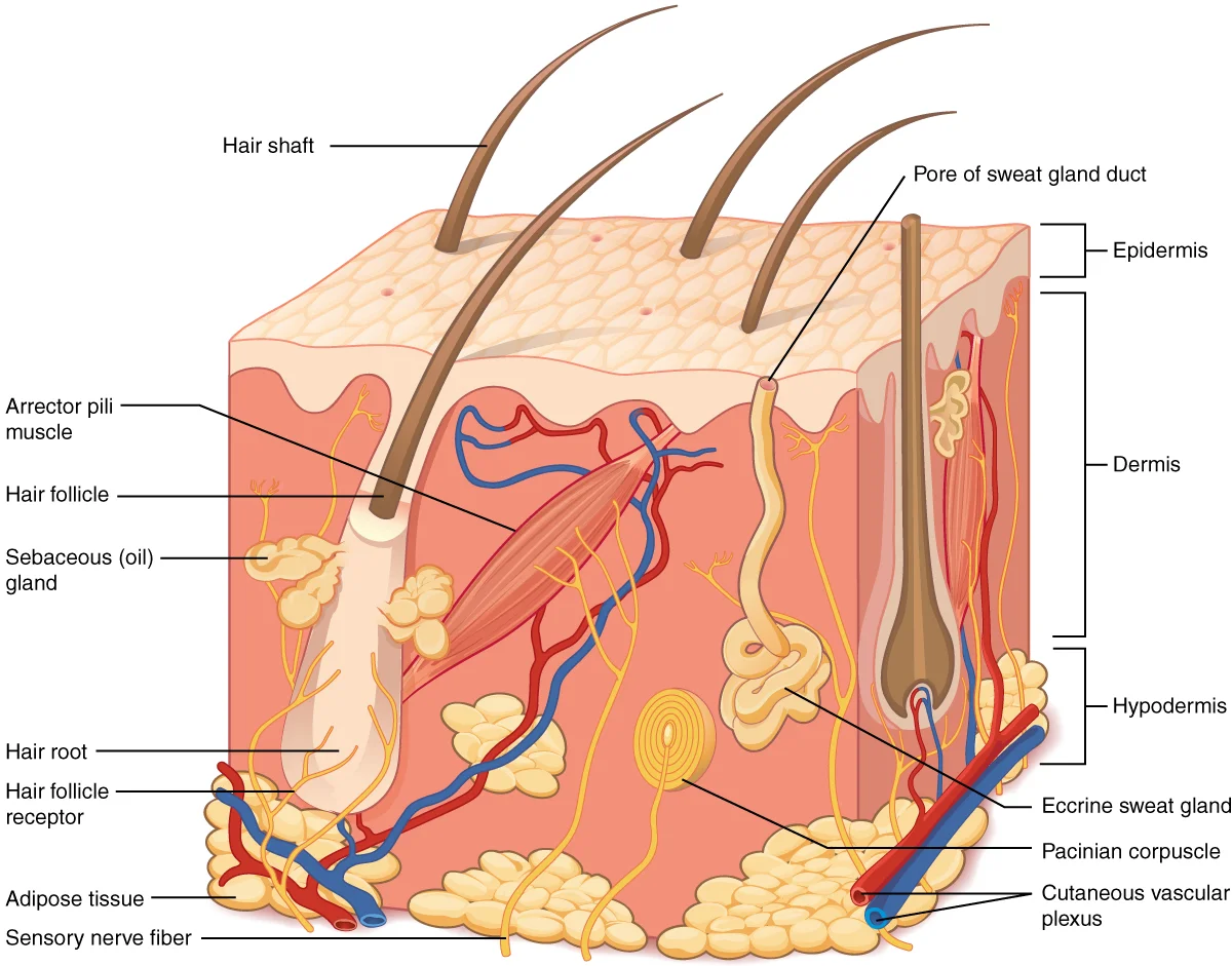
| Epidermis | Stratified squamous epithelium (keratinized) | Avascular, contains keratinocytes and melanocytes, outer protective layer |
| Dermis | Dense irregular connective tissue | Vascularized, contains nerves, glands, hair follicles, collagen/elastin |
| Hypodermis (subcutaneous layer) | Adipose + loose connective tissue | Not technically part of the skin; insulation, energy storage, cushioning |
The Epidermis
The epidermis is the outermost layer. It is avascular (no blood supply) and composed of multiple layers of keratinocytes - cells that produce the tough protein keratin. As keratinocytes divide at the base and move upward, they gradually fill with keratin, flatten, and die. The outermost layer is composed entirely of dead, keratin-packed cells.
The two layers you need to know for the MCAT:
-
Stratum basale (basal layer) - the deepest layer. Contains a single row of stem cells that divide continuously to replenish the epidermis. Also contains melanocytes (produce the pigment melanin) and is attached to the basement membrane.
-
Stratum corneum - the outermost layer. Composed of 20-30 layers of dead, flattened, keratin-packed cells. This is the primary physical barrier and waterproof layer. These cells are continuously shed and replaced (the entire epidermis turns over roughly every 30 days).
Between these two layers, keratinocytes gradually fill with keratin, lose their organelles, and die as they move from the basal layer toward the surface.
Cells of the Epidermis
| Cell Type | Function |
|---|---|
| Keratinocytes | Produce keratin; form the structural bulk of the epidermis (~90% of epidermal cells) |
| Melanocytes | Produce melanin (pigment that absorbs UV radiation); transfer melanin to surrounding keratinocytes |
The Dermis
The dermis is the thick, structural layer beneath the epidermis. Unlike the epidermis, the dermis is vascularized (has blood vessels) and innervated (has nerves). It is made of dense irregular connective tissue containing collagen and elastin fibers, which give skin its strength and elasticity.
The dermis contains:
- Blood vessels - supply nutrients to the epidermis (via diffusion) and play a major role in thermoregulation
- Nerve endings and sensory receptors - detect touch, pressure, temperature, and pain
- Hair follicles and arrector pili muscles (smooth muscle)
- Sweat glands and sebaceous (oil) glands
Skin Appendages
| Structure | Function | Notes |
|---|---|---|
| Hair follicles | Protection (head), sensation (hair movement triggers nerve endings) | Arrector pili muscles (smooth muscle) cause “goosebumps” |
| Sebaceous (oil) glands | Secrete sebum (oily substance that waterproofs and softens skin/hair) | Associated with hair follicles; holocrine secretion |
| Eccrine sweat glands | Thermoregulation via evaporative cooling | Found over entire body surface; secrete watery sweat directly onto skin surface via pores |
| Apocrine sweat glands | Produce thicker secretion (sweat + proteins/lipids) | Found in axillae and groin; empty into hair follicles; activated at puberty; bacterial breakdown of secretion causes body odor |
| Nails | Protect fingertips; assist with fine manipulation | Made of hard keratin; grow from the nail matrix |
Thermoregulation
The skin is the body’s primary thermoregulatory organ. It uses several mechanisms:
When the body is too HOT:
- Vasodilation of dermal blood vessels brings more warm blood near the surface, radiating heat outward
- Eccrine sweat glands produce sweat; evaporation of water from the skin surface removes heat (evaporative cooling)
When the body is too COLD:
- Vasoconstriction of dermal blood vessels reduces blood flow near the surface, conserving heat in the core
- Arrector pili muscles contract (goosebumps) - minimally effective in humans but traps insulating air in furry animals
- Shivering (controlled by skeletal muscle, not skin) generates heat through rapid, involuntary contractions
Vitamin D Synthesis
The skin initiates vitamin D synthesis. UV-B radiation converts a cholesterol precursor in the epidermis to vitamin D₃. This inactive form is then modified in the liver and again in the kidney to produce the active form, calcitriol. Calcitriol promotes calcium absorption in the intestines - connecting the skin directly to calcium homeostasis and bone health.