Pick up a chicken drumstick next time you eat one. Snap it in half and look inside. You will see a hard, dense outer shell surrounding a spongy, honeycomb-like interior. That is not a design flaw - it is brilliant engineering. The dense outer layer resists bending and compression, while the spongy interior saves weight and houses bone marrow. Your bones use the exact same blueprint.
The Two Types of Bone Tissue
All bones contain two types of tissue working together:
Compact (cortical) bone forms the dense outer shell. Under a microscope, compact bone is organized into cylindrical units called osteons (Haversian systems). Each osteon is built like a tree trunk: concentric rings of bone matrix (called lamellae) surround a central canal (the Haversian canal) that carries blood vessels and nerves. Tiny channels called canaliculi radiate outward from each lacuna (the small pocket where a bone cell sits), connecting neighboring bone cells like a network of underground tunnels.
Spongy (cancellous/trabecular) bone fills the interior. It looks like a honeycomb or scaffold - an open lattice of bony struts called trabeculae. This structure is not random. The trabeculae align along lines of mechanical stress, reinforcing the bone exactly where it needs it most. Spongy bone is lighter than compact bone and contains red bone marrow, where blood cells are produced (hematopoiesis).
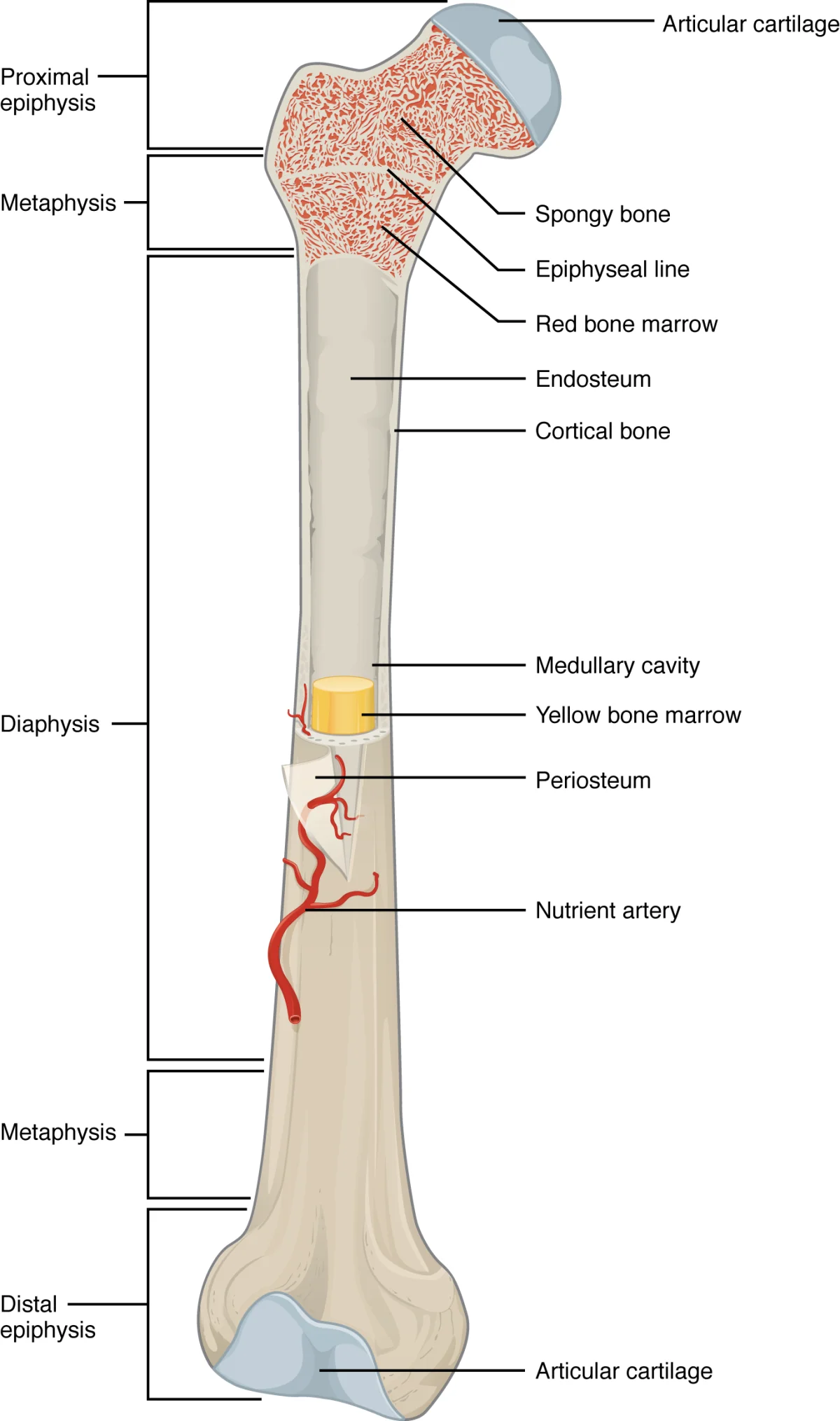
Anatomy of a long bone. The diaphysis (shaft) is a tube of compact bone surrounding the medullary cavity. The epiphyses (ends) contain spongy bone covered by a thin shell of compact bone and capped with articular cartilage. Credit: OpenStax Anatomy and Physiology 2e, CC BY 4.0
Interactive 3D Bone Structure. Rotate to see the periosteum, compact bone, spongy bone, and medullary cavity in cross-section.Credit: zames1992 via Sketchfab, CC BY
Anatomy of a Long Bone
Long bones (like the femur, humerus, and tibia) are the classic model for learning bone anatomy. Here are the key structures:
Structure
Location
Function
Diaphysis
Shaft (middle)
Thick tube of compact bone; encloses the medullary cavity
Epiphysis
Ends (proximal and distal)
Spongy bone covered by thin compact bone; articulates with other bones
Metaphysis
Between diaphysis and epiphysis
Contains the epiphyseal plate (growth plate) in growing bones
Epiphyseal plate
Between metaphysis and epiphysis
Cartilage growth zone; becomes the epiphyseal line when growth stops
Medullary cavity
Interior of diaphysis
Contains yellow bone marrow (fat) in adults
Periosteum
Outer surface (except at joints)
Dense connective tissue; anchors tendons and ligaments; contains osteoblasts for bone repair
Endosteum
Inner surface (lines medullary cavity)
Thin membrane containing osteoblasts and osteoclasts
Articular cartilage
Covers epiphyses at joints
Smooth hyaline cartilage; reduces friction during movement
The Bone Matrix
Bone is not solid mineral. It is a composite material made of two components:
Organic component (~35%) - primarily Type I collagen fibers, plus proteoglycans and glycoproteins. Collagen gives bone its tensile strength and flexibility. Without collagen, bone would be brittle like chalk and shatter on impact.
Inorganic component (~65%) - primarily hydroxyapatite crystals (Ca₁₀(PO₄)₆(OH)₂), a calcium phosphate mineral. Hydroxyapatite gives bone its hardness and compressive strength. Without minerals, bone would be rubbery and bend like cartilage.
This is why bone is both a mineral reservoir and a structural material. When blood calcium drops, the body can dissolve hydroxyapatite to release calcium. When calcium is abundant, it gets deposited back into bone.
The Osteon (Haversian System)
The osteon is the structural and functional unit of compact bone. Understanding its anatomy is essential:
Central (Haversian) canal - runs longitudinally through the center of each osteon; contains blood vessels and nerves
Lamellae - concentric rings of calcified bone matrix surrounding the central canal
Lacunae - small pockets between lamellae where osteocytes (mature bone cells) reside
Canaliculi - tiny channels radiating from each lacuna, forming a network that connects neighboring osteocytes; these allow nutrient exchange and cell-to-cell communication
The Two Skeletal Divisions
The human skeleton is divided into two parts:
Axial skeleton (80 bones) - the central axis of the body: skull, vertebral column (spine), rib cage, and hyoid bone. Provides protection for the brain, spinal cord, and thoracic organs.
Appendicular skeleton (126 bones) - the limbs and the girdles that attach them to the axial skeleton: pectoral girdle (clavicle + scapula), upper limbs, pelvic girdle (hip bones), and lower limbs. Provides the framework for movement.
Embryological Origin
Bones, cartilage, and connective tissues of the musculoskeletal system are derived from the mesoderm, one of the three primary germ layers.
What is the difference between compact bone and spongy bone in terms of structure and location?
Click to reveal answer
Compact bone is dense, organized into osteons (Haversian systems), and forms the outer shell of all bones and the shaft (diaphysis) of long bones. Spongy bone has an open lattice of trabeculae, contains red bone marrow, and is found in the interior of bones, especially at the epiphyses (ends) of long bones. Compact bone provides strength; spongy bone reduces weight and produces blood cells.
A patient has a genetic disorder that produces defective Type I collagen. How would this affect their bones?
Click to reveal answer
The bones would be brittle and fracture easily (this describes osteogenesis imperfecta, or "brittle bone disease"). Collagen provides the organic, flexible component of bone matrix that resists tension. Without functional collagen, the bones retain their mineral hardness but lose their ability to absorb impact - like a ceramic plate that shatters when dropped.
Bone looks like a dead, unchanging material, but it is one of the most metabolically active tissues in your body. Right now, roughly 10% of your skeleton is being torn apart and rebuilt. Three types of cells run this continuous renovation project, and knowing what each one does is absolutely critical for the MCAT.
Osteoblasts - The Builders
Osteoblasts are the bone-forming cells. They synthesize and secrete the organic components of the bone matrix - primarily Type I collagen - in an unmineralized form called osteoid. Once the osteoid is laid down, osteoblasts promote its mineralization by depositing calcium and phosphate crystals (hydroxyapatite).
Key facts about osteoblasts:
Derived from mesenchymal stem cells (the same lineage that produces fibroblasts, chondrocytes, and adipocytes)
Found on the surfaces of bone (periosteum and endosteum)
Once an osteoblast becomes completely surrounded by the matrix it secreted, it becomes trapped and differentiates into an osteocyte
Regulated by parathyroid hormone (PTH), growth hormone, vitamin D, and mechanical stress
Osteoclasts - The Demolition Crew
Osteoclasts are large, multinucleated cells that break down (resorb) bone tissue. They are the demolition crew of the skeletal system.
Key facts about osteoclasts:
Derived from monocyte/macrophage lineage (hematopoietic stem cells) - they are essentially fused macrophages
Multinucleated (formed by fusion of multiple monocyte precursors)
Attach to bone surface and create a sealed resorption pit
Secrete hydrochloric acid (HCl) to dissolve the mineral component and lysosomal enzymes to digest the collagen
The dissolved calcium and phosphate are released into the blood
Stimulated by PTH (indirectly - PTH acts on osteoblasts, which then signal osteoclasts to become active)
Inhibited by calcitonin (from thyroid C cells)
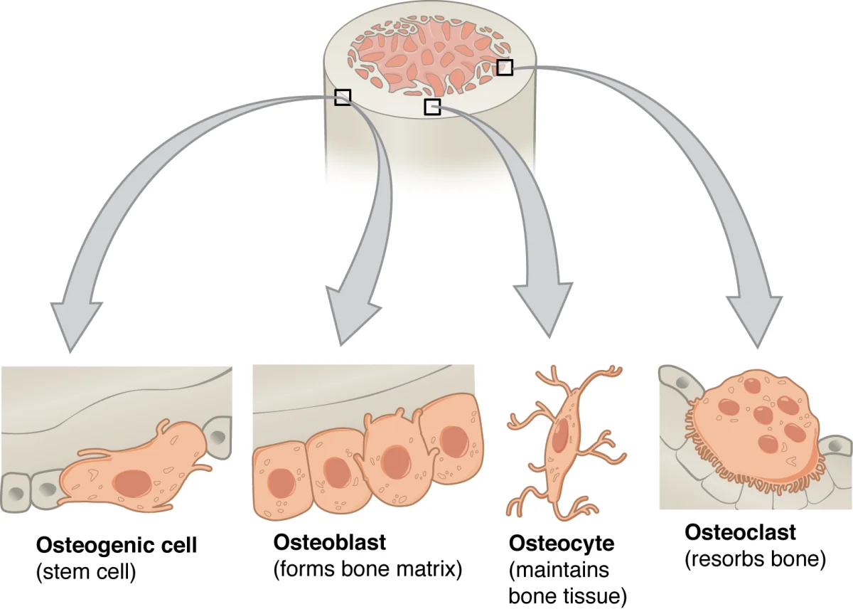
The three types of bone cells. Osteoblasts line the bone surface and deposit new matrix. Osteocytes are mature cells trapped within lacunae and connected by canaliculi. The large, multinucleated osteoclast sits in a resorption pit and breaks down bone. Credit: OpenStax Anatomy and Physiology 2e, CC BY 4.0
Osteocytes - The Maintenance Crew
Osteocytes are the most abundant bone cells (~90-95% of all bone cells). They are mature osteoblasts that have become embedded in the mineralized matrix.
Key facts about osteocytes:
Live inside lacunae (small cavities in the bone matrix)
Extend long cytoplasmic processes through canaliculi to communicate with neighboring osteocytes and cells on the bone surface
Act as mechanosensors - they detect mechanical stress (like the impact of running) and signal osteoblasts to strengthen bone in areas under high stress
Regulate bone remodeling by signaling both osteoblasts and osteoclasts
Can release calcium from the surrounding matrix in response to hormonal signals
How Osteoblasts Control Osteoclasts
An important concept: osteoblasts regulate osteoclast activity. PTH does not directly activate osteoclasts. Instead, PTH binds to receptors on osteoblasts, which then send chemical signals that stimulate osteoclast precursors to mature and begin resorbing bone. So PTH raises blood calcium indirectly, through osteoblasts.
Hormonal Regulation of Bone Cells
The activity of bone cells is tightly regulated by hormones, and the MCAT expects you to know the major players:
Hormone
Source
Effect on Bone
PTH
Parathyroid glands
Stimulates bone resorption (raises blood Ca²⁺)
Calcitonin
Thyroid C cells
Inhibits osteoclasts (lowers blood Ca²⁺)
Vitamin D (calcitriol)
Kidney (activated form)
Promotes Ca²⁺ absorption from gut; supports mineralization
Estrogen
Ovaries
Protects bone; inhibits osteoclast activity
Growth hormone
Anterior pituitary
Stimulates osteoblast activity and bone growth
What cell lineage are osteoclasts derived from, and why is this significant?
Click to reveal answer
Osteoclasts are derived from the monocyte/macrophage lineage (hematopoietic stem cells), NOT from the same mesenchymal lineage as osteoblasts. This is significant because osteoclasts are essentially fused macrophages, which explains their ability to secrete acid and lysosomal enzymes to digest bone - the same tools macrophages use to destroy pathogens.
A patient has chronically elevated PTH levels (primary hyperparathyroidism). What happens to their bone density and blood calcium?
Click to reveal answer
Bone density decreases and blood calcium increases. Chronic PTH elevation causes osteoblasts to continuously signal osteoclasts to become active. The osteoclasts resorb bone faster than osteoblasts can rebuild it, releasing calcium and phosphate into the blood. This leads to hypercalcemia and increased fracture risk.
You were not born with the same skeleton you have now. Most of your bones started as cartilage models that were gradually replaced by bone tissue - a process that began in the womb and was not fully complete until your mid-twenties. Understanding how bone forms and grows is a reliable MCAT topic, and there are two fundamentally different pathways.
Two Pathways of Bone Formation
Intramembranous ossification and endochondral ossification are the two mechanisms the body uses to create bone. They differ in their starting material:
Feature
Intramembranous
Endochondral
Starting material
Mesenchymal connective tissue (membrane)
Hyaline cartilage model
Bones formed
Flat bones of the skull, clavicle, mandible
Most other bones (long bones, vertebrae, pelvis, ribs)
Cartilage intermediate?
No
Yes
Growth plate involvement?
No
Yes (epiphyseal plate)
Intramembranous Ossification
This is the simpler pathway. It produces the flat bones of the skull (frontal, parietal, occipital), the mandible, and the clavicle.
Steps:
Mesenchymal cells cluster together and differentiate directly into osteoblasts
Osteoblasts secrete osteoid (unmineralized bone matrix), which then mineralizes
The mineralized matrix forms small islands called trabeculae, which grow and merge
Blood vessels are incorporated between the trabeculae
The outer surface condenses into a layer of compact bone; the interior remains spongy bone
The periosteum forms from the surrounding mesenchyme
The key point: there is no cartilage intermediate. Mesenchyme transforms directly into bone.
Endochondral Ossification
This is the pathway used by most bones in the body, including all long bones. It starts with a cartilage “blueprint” that is systematically replaced by bone.
Steps of endochondral ossification:
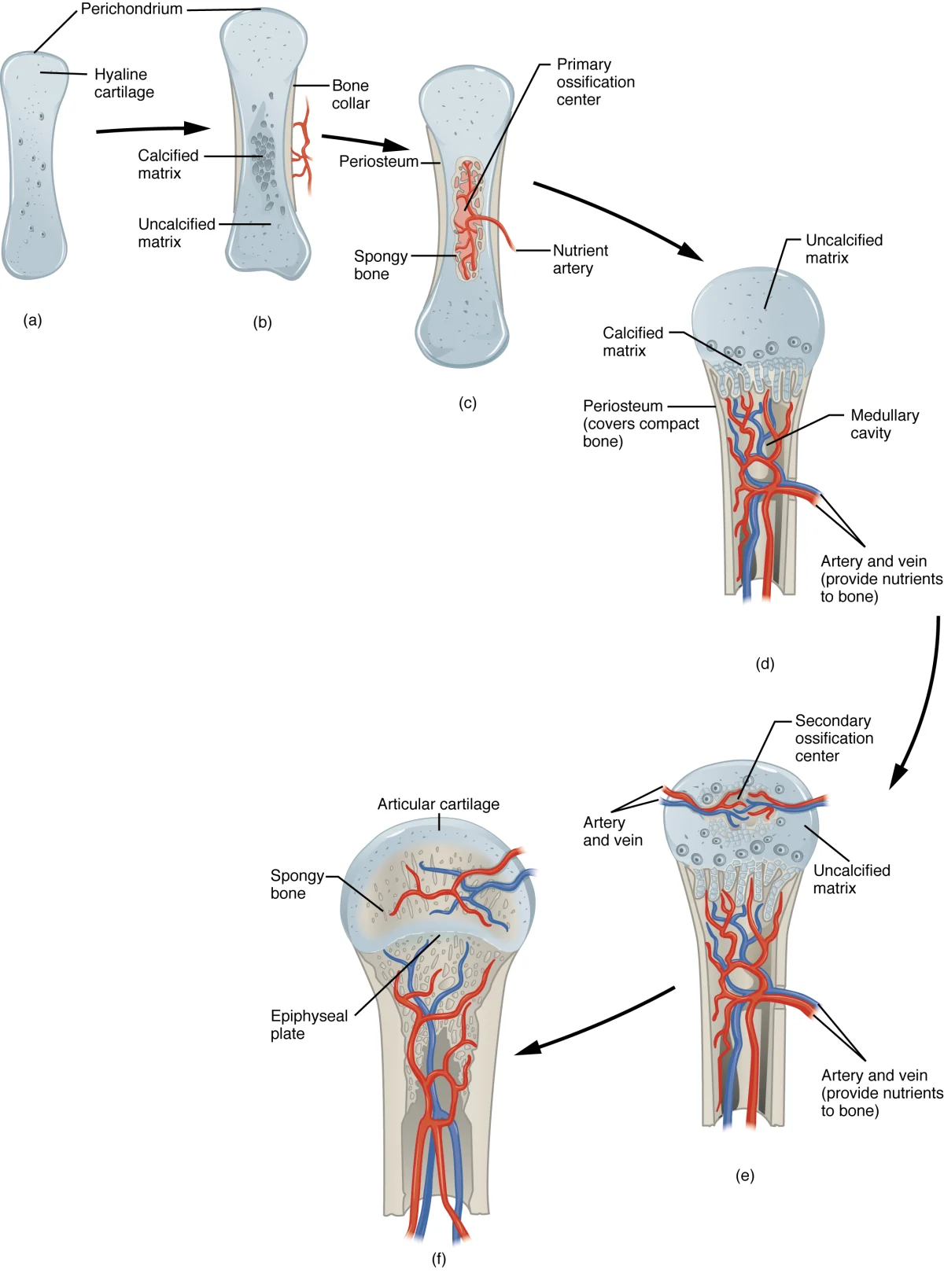
Cartilage model forms - Mesenchymal cells differentiate into chondrocytes, which secrete a hyaline cartilage model in the shape of the future bone
Primary ossification center - In the center of the diaphysis (shaft), chondrocytes enlarge (hypertrophy), calcify the surrounding cartilage, and then die. Blood vessels invade the area, bringing osteoblasts that begin depositing true bone on the calcified cartilage scaffolding
Bone collar forms - Osteoblasts in the perichondrium (the membrane surrounding the cartilage) lay down a collar of compact bone around the diaphysis, converting the perichondrium into the periosteum
Medullary cavity forms - Osteoclasts break down the newly formed spongy bone in the center of the diaphysis, creating the medullary cavity
Secondary ossification centers - Blood vessels invade the epiphyses (ends), and the same process of cartilage replacement occurs there. However, spongy bone remains in the epiphyses (it is not hollowed out like the diaphysis)
Epiphyseal plate remains - A band of cartilage persists between the diaphysis and each epiphysis - this is the growth plate
Stages of endochondral ossification. The process begins with a cartilage model (left), followed by formation of the bone collar and primary ossification center, invasion of blood vessels, development of the medullary cavity, and finally appearance of secondary ossification centers in the epiphyses. Credit: OpenStax Anatomy and Physiology 2e, CC BY 4.0
The Epiphyseal (Growth) Plate
The epiphyseal plate is the engine of longitudinal bone growth. It is a disc of hyaline cartilage located between the epiphysis and diaphysis. The basic process is straightforward:
On the epiphyseal side, chondrocytes actively divide and produce new cartilage (pushing the plate outward)
On the diaphyseal side, older cartilage calcifies, the chondrocytes die, and osteoblasts replace the cartilage with bone
This means new cartilage is constantly being added on one side and replaced by bone on the other. The net result: the bone gets longer as the epiphysis is pushed farther from the diaphysis.
Bone Remodeling
Even after growth is complete, bone is continuously remodeled throughout life. Remodeling is the coordinated process of bone resorption by osteoclasts followed by new bone deposition by osteoblasts. About 10% of the adult skeleton is replaced each year.
Wolff’s law states that bone remodels in response to mechanical stress placed upon it. Bone is deposited where stress is high and resorbed where stress is low. This explains why:
Weight-bearing exercise increases bone density
Astronauts lose bone mass in microgravity (no mechanical loading)
A bedridden patient develops osteoporosis in immobilized limbs
The dominant arm of a tennis player has denser bones than the non-dominant arm
The Calcium Axis: Bone Meets Endocrine
Remodeling is the skeleton’s role in calcium homeostasis. Three hormones work together to keep blood Ca²⁺ in range:
PTH (parathyroid) rises when blood Ca²⁺ is low. It stimulates osteoclast-driven resorption (via osteoblasts), boosts Ca²⁺ reabsorption in the kidney, and activates vitamin D.
Calcitriol (active vitamin D, made in the kidney) increases gut Ca²⁺ absorption and supports mineralization.
Calcitonin (thyroid C cells) opposes PTH: it inhibits osteoclasts, lowering blood Ca²⁺.
Fracture Repair
When a bone breaks, the repair process recapitulates many of the steps of endochondral ossification:
Hematoma formation - Blood from ruptured vessels clots at the fracture site
Fibrocartilaginous callus - Fibroblasts and chondrocytes invade the clot and produce a soft callus of fibrocartilage that bridges the gap
Bony (hard) callus - Osteoblasts replace the fibrocartilage with spongy bone (endochondral ossification in miniature)
Remodeling - Osteoclasts reshape the bony callus into compact bone, restoring the original bone structure over months to years
What is the difference between intramembranous and endochondral ossification?
Click to reveal answer
Intramembranous ossification forms bone directly from mesenchymal connective tissue with no cartilage intermediate (produces flat bones of the skull and clavicle). Endochondral ossification replaces a pre-existing hyaline cartilage model with bone tissue (produces most other bones, including all long bones). Both use osteoblasts to deposit bone matrix, but only endochondral ossification involves a cartilage template.
An astronaut returns from 6 months on the International Space Station. What has happened to their bone density, and why?
Click to reveal answer
Their bone density has decreased. According to Wolff's law, bone remodels in response to mechanical stress. In microgravity, there is minimal mechanical loading on the skeleton, so osteoclast resorption continues but osteoblast deposition slows. The result is net bone loss, similar to the osteoporosis seen in bedridden patients. Astronauts can lose 1-2% of bone mass per month in space.
Bend your ear forward. It springs back. Now press on the tip of your nose - it squishes and returns to shape. That flexibility comes from cartilage, a specialized connective tissue that is softer and more flexible than bone but firmer than most other tissues. There are three types of cartilage in your body, and they each have a different job based on their specific combination of collagen and elastic fibers.
General Properties of Cartilage
Before diving into the three types, know these features that ALL cartilage shares:
Avascular - cartilage has no blood supply. Nutrients reach chondrocytes by diffusing through the matrix from surrounding tissues. This is why cartilage heals slowly (or not at all) after injury.
No nerves - cartilage is not innervated (aneural), which is why you can press on your ear without pain
Chondrocytes - the only cell type in cartilage; they sit in small cavities called lacunae and produce the surrounding matrix
Perichondrium - a dense connective tissue membrane covering most cartilage (except articular cartilage and fibrocartilage). It provides blood supply to the outer edge and contains stem cells for cartilage growth
High water content - the matrix is heavily hydrated, giving cartilage its ability to resist compression
Hyaline Cartilage
Hyaline cartilage is the most common type. Its name comes from the Greek word “hyalos” (glass) because it appears glassy and translucent when fresh.
Matrix composition: Fine Type II collagen fibers in a gel-like ground substance rich in proteoglycans (especially aggrecan). The collagen fibers are too thin to see individually, giving the matrix its smooth, glassy appearance.
Key locations:
Articular surfaces of joints (covering bone ends - no perichondrium here)
Tracheal rings and bronchi (keeps airways open)
Tip of the nose
Costal cartilage (connects ribs to sternum)
Epiphyseal plates (growth plates in growing bones)
Fetal skeleton (the template for endochondral ossification)
Function: Provides smooth, low-friction surfaces at joints; provides flexible support for airways; serves as the template for bone growth.
Elastic Cartilage
Elastic cartilage is similar to hyaline cartilage but contains a dense network of elastic fibers in addition to Type II collagen. This makes it extremely flexible - it can be bent repeatedly without breaking.
Key locations:
External ear (pinna/auricle)
Epiglottis (the flap that covers the larynx during swallowing)
Auditory (Eustachian) tube
Function: Provides flexible support that can spring back to its original shape after being deformed.
Fibrocartilage
Fibrocartilage is the toughest type. It contains dense bundles of Type I collagen fibers (the same collagen found in bone, tendons, and ligaments), making it extremely resistant to compression and tension.
Key locations:
Intervertebral discs (between vertebrae)
Menisci of the knee
Pubic symphysis
Labrum of the hip and shoulder joints
At the insertion points of tendons and ligaments into bone
Function: Absorbs compressive shock and resists tearing at sites that experience heavy mechanical stress.
Important: Fibrocartilage has no perichondrium (unlike hyaline and elastic cartilage). Its chondrocytes are scattered among thick collagen bundles rather than enclosed in a smooth matrix.
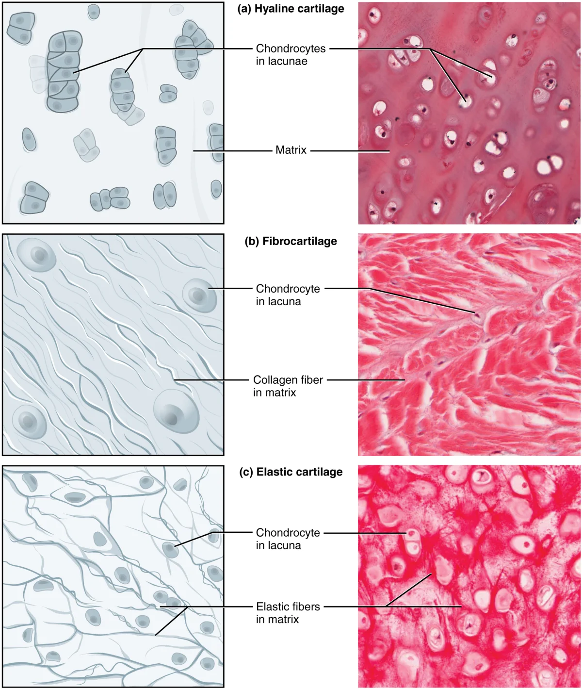
The three types of cartilage. Hyaline cartilage (left) has a smooth, glassy matrix. Elastic cartilage (center) contains additional elastic fibers for flexibility. Fibrocartilage (right) has dense Type I collagen bundles for strength. Credit: OpenStax Anatomy and Physiology 2e, CC BY 4.0
Comparison Table
Feature
Hyaline
Elastic
Fibrocartilage
Collagen type
Type II
Type II + elastic fibers
Type I (thick bundles)
Flexibility
Moderate
Very high
Low
Strength
Moderate
Moderate
Very high
Perichondrium?
Yes (except articular)
Yes
No
Key locations
Trachea, joints, nose, growth plates
Ear, epiglottis
Intervertebral discs, meniscus, pubic symphysis
Appearance
Glassy, translucent
Yellow, flexible
Dense, fibrous
Cartilage Growth
Cartilage can grow in two ways:
Interstitial growth - chondrocytes within the cartilage divide and produce new matrix from the inside. This occurs mainly during childhood and at the epiphyseal plate.
Appositional growth - new cartilage is added to the surface by cells in the perichondrium that differentiate into chondrocytes. This can occur throughout life.
Why does cartilage heal slowly after injury?
Click to reveal answer
Cartilage is avascular (has no blood supply). Without blood vessels, inflammatory cells, growth factors, and nutrients cannot be efficiently delivered to the injury site. Bone, by contrast, has an extensive blood supply and heals much more effectively. This is why torn meniscus cartilage in the knee often requires surgical intervention.
Which type of cartilage is found in the intervertebral discs, and what makes it suited for this location?
Click to reveal answer
Fibrocartilage. It contains thick bundles of Type I collagen, making it the toughest cartilage type. The intervertebral discs must absorb compressive forces from body weight and movement, and fibrocartilage's combination of strength and shock absorption makes it ideal for this role. It also lacks a perichondrium, distinguishing it from hyaline and elastic cartilage.
Bones are rigid. If your skeleton were a single solid piece, you could not move at all. Joints are the points where two or more bones meet, and they range from completely immovable (like the sutures in your skull) to highly mobile (like your shoulder). The type of joint determines how much movement is possible.
Joint Classification by Structure
Joints are classified into three structural categories based on the type of tissue connecting the bones:
Diarthrosis - freely movable (e.g., all synovial joints)
The Synovial Joint
Synovial joints are the most common and most tested type on the MCAT. They share a characteristic set of structures:
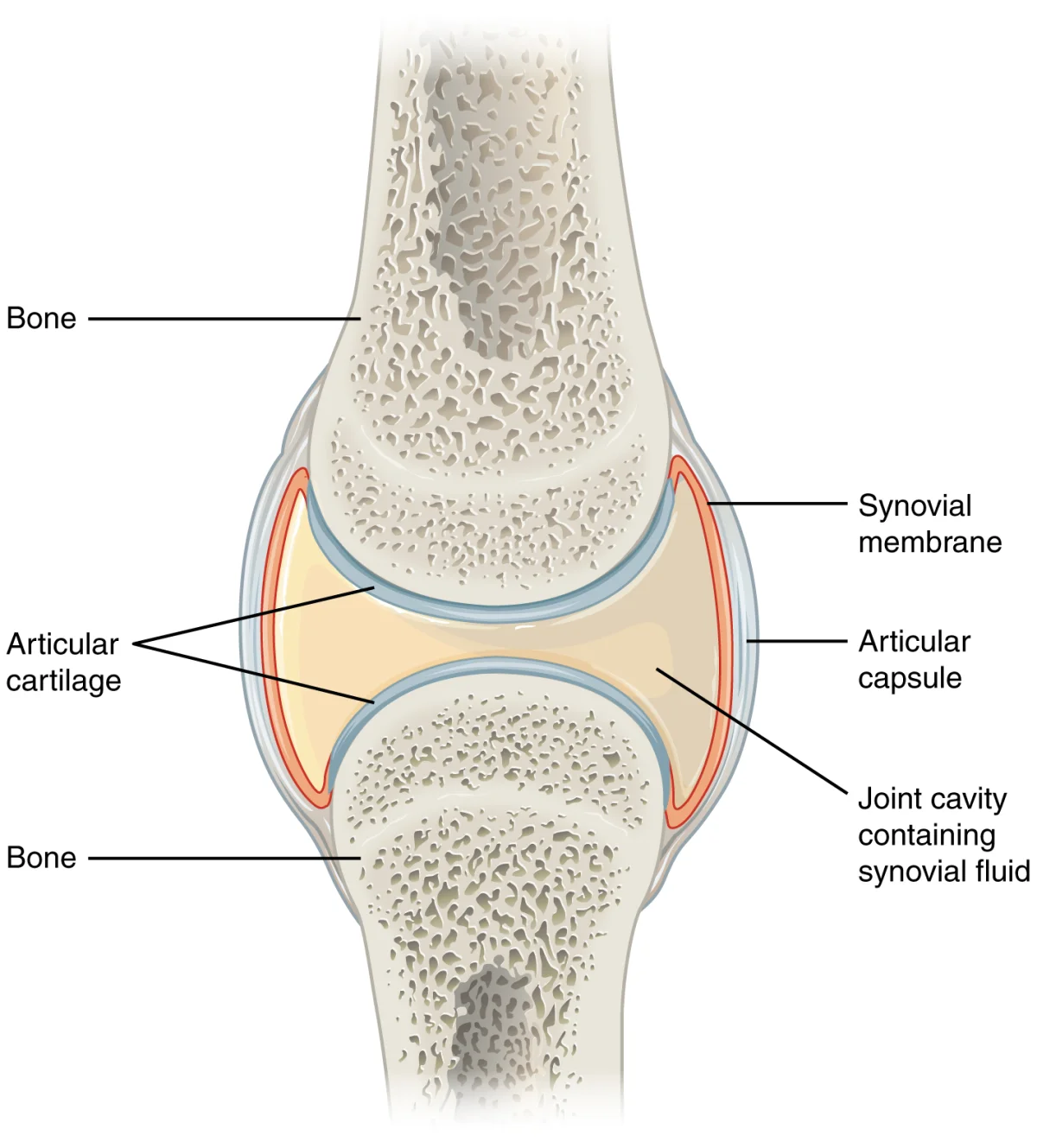
Anatomy of a synovial joint. The articular cartilage covers the bone ends, the synovial membrane secretes synovial fluid into the joint cavity, and the fibrous capsule provides structural support. Ligaments reinforce the capsule. Credit: OpenStax Anatomy and Physiology 2e, CC BY 4.0
Interactive 3D Synovial Joints. Explore the six types of synovial joints and how their shapes determine the range of motion they allow.Credit: greglittle via Sketchfab, CC BY
Key components of a synovial joint:
Articular (hyaline) cartilage - smooth cartilage covering the bone ends; reduces friction and absorbs shock. Has no perichondrium and is avascular.
Joint (synovial) cavity - the space between the bones, filled with synovial fluid
Synovial membrane - lines the inner surface of the joint capsule (but NOT the articular cartilage). Secretes synovial fluid.
Synovial fluid - viscous fluid that lubricates the joint surfaces and nourishes the avascular articular cartilage. Contains hyaluronic acid (for viscosity) and filtered plasma (for nutrients).
Fibrous capsule - tough outer layer of the joint capsule made of dense connective tissue. Reinforced by ligaments.
Ligaments - bands of dense connective tissue connecting bone to bone. They stabilize the joint and limit excessive movement.
Bursae - small, fluid-filled sacs that reduce friction between tendons, ligaments, and bones at points of high mechanical stress
Types of Synovial Joints
The three synovial joint types worth knowing for the MCAT:
Type
Movement
Example
Hinge
Flexion/extension (1 axis)
Elbow, knee
Ball-and-socket
Movement in all directions (3 axes)
Shoulder, hip
Pivot
Rotation (1 axis)
Atlas/axis (turning the head)
Tendons and Ligaments
Tendons and ligaments are both made of dense regular connective tissue (primarily Type I collagen fibers arranged in parallel bundles), but they connect different structures:
Feature
Tendons
Ligaments
Connects
Muscle to bone
Bone to bone
Function
Transmits muscle force to bone to produce movement
Stabilizes joints, limits excessive movement
Vascularity
Relatively avascular
Relatively avascular
Healing
Slow (poor blood supply)
Slow (poor blood supply)
Elasticity
Low (stiff - efficient force transfer)
Slightly more elastic than tendons
Example
Achilles tendon, rotator cuff tendons
ACL (anterior cruciate ligament), MCL
Movement Terminology
The MCAT expects you to recognize the basic types of joint movements:
Movement
Description
Example
Flexion
Decreasing the angle between bones
Bending the elbow, bending the knee
Extension
Increasing the angle between bones
Straightening the elbow, straightening the knee
Abduction
Moving a limb away from the body’s midline
Raising your arm out to the side
Adduction
Moving a limb toward the body’s midline
Lowering a raised arm back to your side
Rotation
Turning a bone around its own axis
Turning your head left or right
Circumduction
Circular movement combining flexion, extension, abduction, and adduction
Swinging your arm in a circle
Antagonistic and Synergistic Muscles
Muscles work in coordinated pairs and groups:
Antagonistic muscles produce movement in opposite directions. When one contracts, the other relaxes. Example: the biceps (flexor) and triceps (extensor) at the elbow. When the biceps contracts and flexes the elbow, the triceps relaxes. When the triceps contracts and extends the elbow, the biceps relaxes.
Synergistic muscles assist each other to produce the same movement. Example: the hamstring muscles (biceps femoris, semitendinosus, semimembranosus) all work together to flex the knee.
Agonist (prime mover) - the muscle primarily responsible for a movement
Antagonist - the muscle that opposes the agonist
Synergist - a muscle that assists the agonist
Other Connective Tissues
A few additional structures are worth knowing:
Aponeurosis - a flat, sheet-like tendon (e.g., the aponeurosis connecting the abdominal muscles)
Fascia - dense connective tissue that surrounds muscles, groups of muscles, blood vessels, and nerves. It compartmentalizes structures and reduces friction.
Meniscus - a crescent-shaped pad of fibrocartilage in certain joints (knee, temporomandibular joint) that acts as a shock absorber and improves joint fit
What are the key structural differences between a fibrous joint, a cartilaginous joint, and a synovial joint?
Click to reveal answer
Fibrous joints are connected by dense connective tissue (collagen fibers), have no joint cavity, and are mostly immovable (e.g., skull sutures). Cartilaginous joints are connected by cartilage (hyaline or fibrocartilage), have no joint cavity, and are slightly movable (e.g., intervertebral discs). Synovial joints have a fluid-filled joint cavity lined by a synovial membrane, are surrounded by a fibrous capsule, and are freely movable (e.g., knee, shoulder).
What is the function of synovial fluid, and what produces it?
Click to reveal answer
Synovial fluid is secreted by the synovial membrane lining the joint capsule. It serves two functions: (1) lubrication - it reduces friction between articular cartilage surfaces during movement, and (2) nutrition - it delivers nutrients to and removes waste from the avascular articular cartilage through diffusion. It contains hyaluronic acid (giving it viscosity) and filtered plasma components.
Before we can understand how muscles contract, we need to understand what they are made of. Skeletal muscle has an elegant, repeating architecture that is organized like a set of Russian nesting dolls - each level is a smaller version of the one above it.
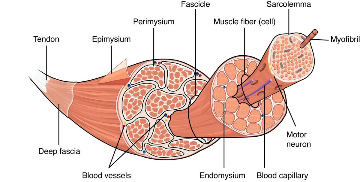
The Muscle Hierarchy
From largest to smallest:
Level
What It Is
Wrapped In
Muscle
The whole organ (e.g., biceps brachii)
Epimysium (dense connective tissue)
Fascicle
A bundle of muscle fibers
Perimysium
Muscle fiber
A single multinucleated cell
Endomysium
Myofibril
A contractile thread running the length of the fiber
Sarcolemma encloses all myofibrils
Sarcomere
The functional unit of contraction
Bounded by Z lines
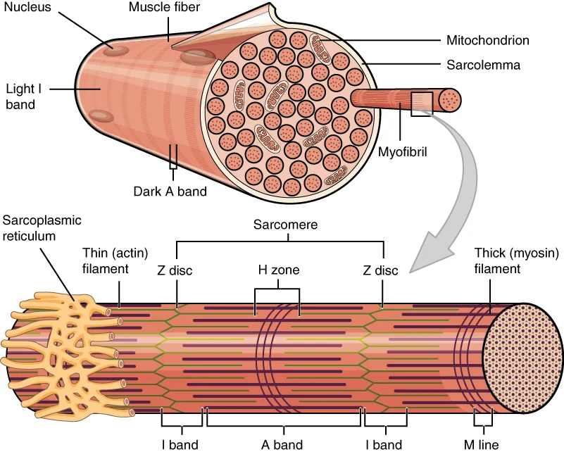
Skeletal muscle hierarchy. Focus on: muscle to fascicle to muscle fiber to myofibril to sarcomere. Know that epimysium, perimysium, and endomysium wrap each level, but the MCAT mostly cares about the sarcomere level and below. Credit: OpenStax Anatomy and Physiology 2e, CC BY 4.0
Key Features of a Muscle Fiber (Cell)
A single skeletal muscle fiber is one of the largest and most unusual cells in the body:
Multinucleated - each fiber contains dozens to hundreds of nuclei, pushed to the periphery of the cell. This is because muscle fibers form by the fusion of many precursor cells (myoblasts) during development.
Sarcolemma - the cell membrane of a muscle fiber
Sarcoplasm - the cytoplasm of a muscle fiber
Sarcoplasmic reticulum (SR) - a specialized smooth endoplasmic reticulum that stores and releases Ca²⁺. This is the calcium reservoir that triggers contraction.
T-tubules (transverse tubules) - invaginations of the sarcolemma that plunge deep into the fiber. They carry the action potential from the surface to the interior, ensuring the entire fiber contracts simultaneously.
Triad - the functional unit of excitation-contraction coupling: one T-tubule flanked by two terminal cisternae of the SR. This arrangement ensures that the electrical signal (T-tubule) is right next to the calcium store (SR).
The Sarcomere - The Functional Unit of Contraction
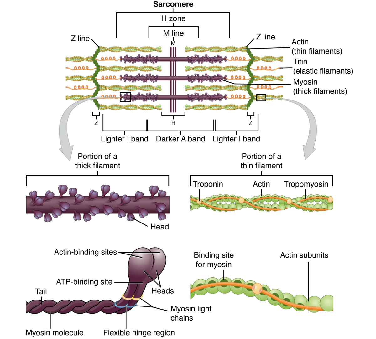
The sarcomere is the repeating unit that makes muscle contraction possible. Each myofibril is a chain of thousands of sarcomeres arranged end to end. The sarcomere is defined as the region between two Z lines (also called Z discs).
Structure of the sarcomere. Thin (actin) filaments are anchored to the Z lines. Thick (myosin) filaments span the A band. The H zone and I band change width during contraction, but the A band stays the same. Credit: OpenStax Anatomy and Physiology 2e, CC BY 4.0
Sarcomere Components
The two main filaments:
Component
Thick Filament
Thin Filament
Main protein
Myosin (with ATPase heads)
Actin (globular subunits forming F-actin)
Regulatory proteins
None on the filament itself
Tropomyosin (covers myosin-binding sites on actin) and Troponin (Ca²⁺ sensor)
Location
Center of sarcomere
Anchored to Z lines, extend toward center
Diameter
~15 nm (thick)
~7 nm (thin)
The bands and zones:
Structure
What It Contains
Appearance
Changes During Contraction?
A band
Entire length of thick (myosin) filaments, including any overlap with thin filaments
Dark
NO - stays the same width (A = Always the same)
I band
Thin (actin) filaments ONLY (no thick filament overlap)
Light
YES - gets SHORTER
H zone
Thick (myosin) filaments ONLY (no thin filament overlap)
Light region within A band
YES - gets SHORTER
Z line (Z disc)
Protein disc anchoring thin filaments
Thin dark line
Moves CLOSER to center during contraction
M line
Proteins connecting thick filaments at the center
Thin line in center of H zone
Stays in center
Regulatory Proteins: Troponin and Tropomyosin
These two proteins on the thin filament act as the molecular “safety lock” that prevents contraction when the muscle should be relaxed:
Tropomyosin - a long, thread-like protein that wraps around the actin filament, physically covering the myosin-binding sites. When tropomyosin is in the blocking position, myosin heads cannot attach to actin, and contraction cannot occur.
Troponin - a regulatory complex that sits on tropomyosin at regular intervals. The key subunit is troponin C, which binds calcium (Ca²⁺). When Ca²⁺ binds to troponin C, the troponin complex changes shape, pulling tropomyosin away from the myosin-binding sites on actin. This exposes the binding sites and allows cross-bridge cycling (contraction) to begin.
During muscle contraction, which sarcomere bands/zones change in width and which stay the same?
Click to reveal answer
The I band and H zone get shorter during contraction (because thin filaments slide further over thick filaments, increasing overlap). The A band stays the same width because it spans the entire length of the thick filaments, which do not change length. The Z lines move closer together, shortening the sarcomere overall. Remember: A band = Always the same.
What is the role of troponin C in muscle contraction?
Click to reveal answer
Troponin C binds calcium ions (Ca²⁺). When Ca²⁺ is released from the sarcoplasmic reticulum, it binds to troponin C, causing a conformational change in the troponin complex. This pulls tropomyosin away from the myosin-binding sites on actin, exposing them and allowing myosin heads to form cross-bridges. Without Ca²⁺ binding to troponin C, tropomyosin blocks the binding sites and the muscle remains relaxed.
Here is the central question of muscle physiology: how does a muscle generate force? The answer is the sliding filament model - the most important concept in this entire chapter. The filaments themselves do not shorten. Instead, the thin filaments slide over the thick filaments, pulling the Z lines closer together and shortening the sarcomere. Multiply this across millions of sarcomeres contracting simultaneously, and you get the gross movement of the whole muscle.
Key Principles of the Sliding Filament Model
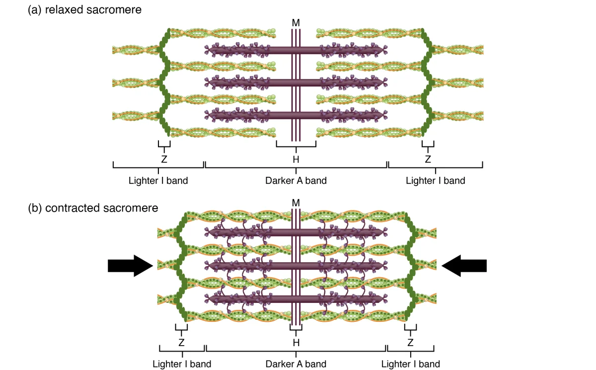
Filaments do NOT shorten. The actin and myosin filaments maintain their length throughout contraction. The sarcomere shortens because the filaments slide past each other.
The A band does not change width. The A band spans the full length of the thick filaments, which do not change length.
The I band and H zone shrink. As thin filaments slide inward over thick filaments, the regions containing only one type of filament get smaller.
Force is generated by cross-bridge cycling - the repeated attachment, pivoting, and detachment of myosin heads on actin filaments.
ATP is required for both contraction AND relaxation. ATP powers the myosin head reset and is required for myosin to release from actin.
The sliding filament model. In the relaxed state (top), the I bands and H zone are wide. During contraction (bottom), the thin filaments slide inward, narrowing the I bands and H zone while the A band remains unchanged. Credit: OpenStax Anatomy and Physiology 2e, CC BY 4.0
Interactive 3D Sarcomere. Rotate to see how thick (myosin) and thin (actin) filaments interlock between Z lines. The filaments slide past each other during contraction.Credit: rgmz2015 via Sketchfab, CC BY
Cross-Bridge Cycling - The Molecular Motor
Cross-bridge cycling is the step-by-step process by which myosin heads generate force on actin. Each cycle produces a small “power stroke” that pulls the thin filament toward the center of the sarcomere.
The Four Steps of Cross-Bridge Cycling
| Step | What Happens | Energy Source | State of Myosin Head |
|---|---|---|---|
| 1. Cross-bridge formation | Myosin head (energized, cocked position) binds to exposed binding site on actin | None (uses energy stored from previous step) | Bound to actin, high-energy conformation |
| 2. Power stroke | Myosin head pivots, pulling the thin filament toward the M line. ADP and Pi are released. | Stored elastic energy (from ATP hydrolysis in step 4) | Bound to actin, low-energy conformation |
| 3. Cross-bridge detachment | A new ATP molecule binds to the myosin head, causing it to release from actin | ATP binding (NOT hydrolysis) | Detached from actin, low-energy |
| 4. Myosin head reset (re-cocking) | Myosin’s ATPase hydrolyzes ATP → ADP + Pi. The energy is used to return the myosin head to its high-energy (cocked) position. | ATP hydrolysis | Detached from actin, high-energy (ready for next cycle) |
Then the cycle repeats from Step 1, as long as Ca²⁺ is present (keeping binding sites on actin exposed) and ATP is available.
The Role of ATP in Contraction
ATP plays TWO critical roles in muscle contraction:
Detachment - ATP binding to the myosin head breaks the cross-bridge (allows myosin to release from actin). Without ATP, myosin remains locked on actin.
Re-cocking - ATP hydrolysis (ATP → ADP + Pi) provides the energy to return the myosin head to its high-energy position, ready for the next power stroke.
Notice that ATP is NOT used during the power stroke itself. The power stroke is powered by the elastic energy that was stored in the myosin head during the previous hydrolysis step. The ATP is used to “reload the spring.”
The Role of Calcium in Contraction
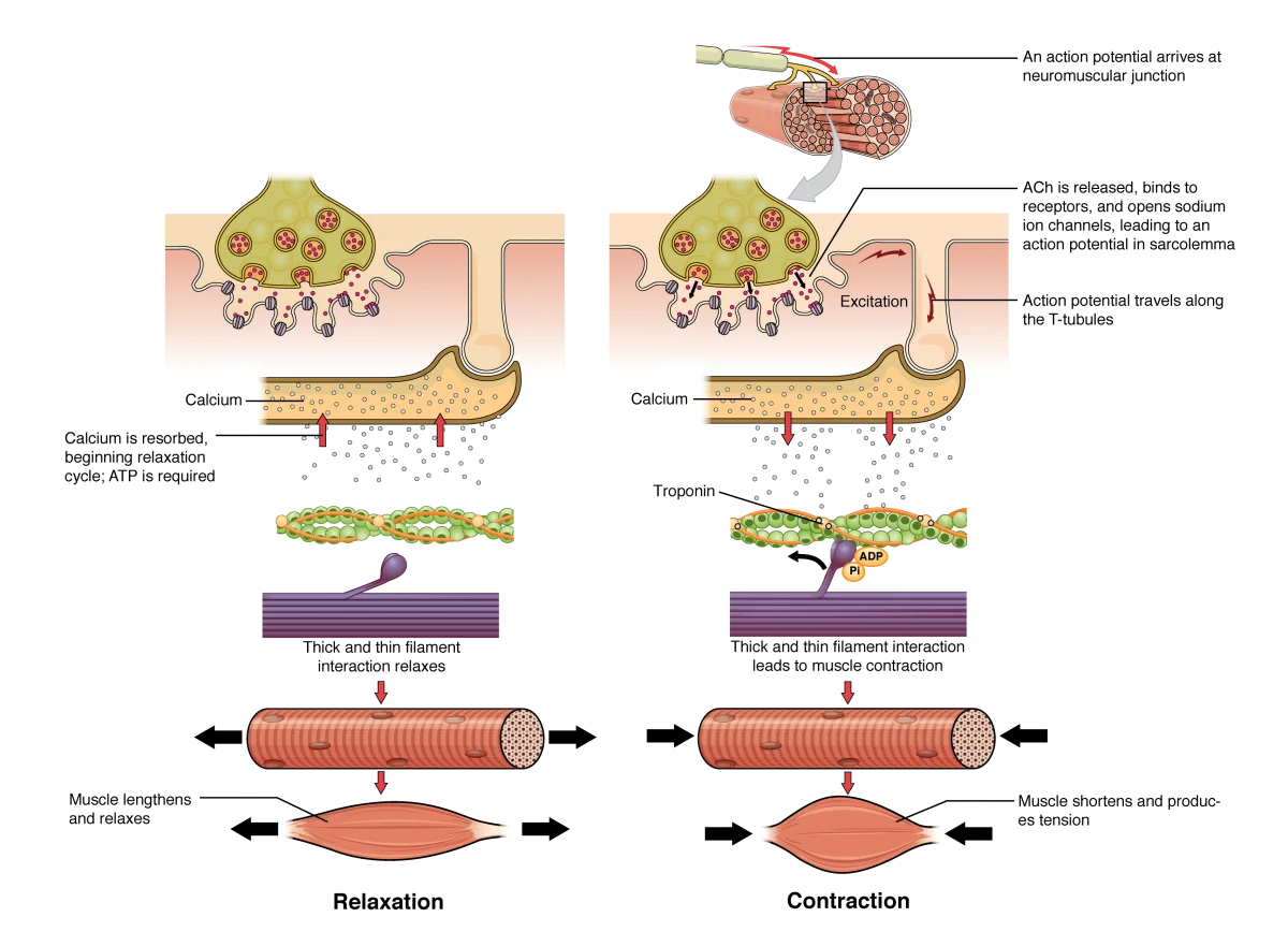
Ca²⁺ is the ON switch for muscle contraction. Here is the sequence:
At rest, tropomyosin covers the myosin-binding sites on actin. Cross-bridges cannot form.
When Ca²⁺ is released from the sarcoplasmic reticulum, it binds to troponin C.
Troponin changes shape, pulling tropomyosin away from the binding sites.
Myosin-binding sites are now exposed, and cross-bridge cycling begins.
When Ca²⁺ is pumped back into the SR (by the SERCA pump, which requires ATP), troponin releases Ca²⁺, tropomyosin slides back over the binding sites, and contraction stops.
The Length-Tension Relationship
The force a sarcomere generates depends on the degree of overlap between thick and thin filaments:
Optimal overlap - maximum cross-bridges can form → maximum force
Overstretched (too little overlap) - fewer cross-bridges can form → reduced force. At extreme stretch, no overlap → zero force.
Over-compressed (too much overlap) - thin filaments from opposite sides interfere with each other, and thick filaments butt against Z lines → reduced force
This produces a bell-shaped length-tension curve. The MCAT may present this curve and ask you to explain why force changes at different sarcomere lengths.
The Force-Velocity Relationship
Force and velocity of contraction have an inverse relationship:
Heavy load, slow contraction - when the load is large, each myosin head takes longer to pull the thin filament. Velocity drops toward zero as load approaches the muscle’s maximum force.
Light load, fast contraction - when the load is small, thin filaments slide quickly past thick filaments. Velocity is highest with no load.
This is why you can lift a book fast but a heavy barbell slowly. The MCAT may show this hyperbolic curve and ask what happens when load changes.
A fresh ATP molecule binds to a myosin head that is attached to actin. What happens immediately?
Click to reveal answer
The myosin head detaches from actin. ATP binding (not hydrolysis) causes a conformational change in the myosin head that breaks the cross-bridge. This is Step 3 of cross-bridge cycling. After detachment, the myosin ATPase hydrolyzes ATP → ADP + Pi, and the energy is used to re-cock the myosin head to its high-energy position (Step 4), preparing it for the next cycle.
Why does muscle contraction require ATP for BOTH contraction and relaxation?
Click to reveal answer
Contraction requires ATP for two steps of cross-bridge cycling: (1) ATP binding detaches myosin from actin, and (2) ATP hydrolysis re-cocks the myosin head. Relaxation requires ATP because the SERCA pump (Ca²⁺-ATPase) on the sarcoplasmic reticulum uses ATP to actively pump Ca²⁺ back into the SR. Without this active transport, Ca²⁺ remains in the cytoplasm, troponin C stays bound to Ca²⁺, and the muscle stays contracted.
You now know the molecular machinery of contraction (cross-bridge cycling) and its on-switch (calcium). But how does a decision in your brain translate into calcium flooding a muscle cell? That is excitation-contraction (E-C) coupling - the chain of events that converts an electrical signal (action potential) into a mechanical response (muscle contraction).
The Complete E-C Coupling Pathway
Follow each step carefully - this sequence is one of the most commonly tested pathways on the MCAT:
Step 1: Action potential arrives at the neuromuscular junction
A motor neuron releases acetylcholine (ACh) into the synaptic cleft. ACh binds to nicotinic receptors on the sarcolemma (muscle cell membrane), opening Na⁺ channels and depolarizing the muscle fiber.
Step 2: Action potential propagates along the sarcolemma
The depolarization triggers a full action potential that spreads across the entire surface of the muscle fiber.
Step 3: Action potential dives into T-tubules
The action potential travels down the T-tubules (transverse tubules) - deep invaginations of the sarcolemma that penetrate into the interior of the fiber. This ensures the signal reaches every sarcomere simultaneously, even those deep inside the cell.
Step 4: Voltage sensor activates calcium release
The T-tubule membrane contains voltage-sensitive proteins called dihydropyridine (DHP) receptors (L-type calcium channels). When the action potential depolarizes the T-tubule, DHP receptors change conformation and physically interact with ryanodine receptors (RyR) on the adjacent sarcoplasmic reticulum membrane.
Step 5: SR releases calcium
Activated ryanodine receptors open, and Ca²⁺ floods out of the SR into the sarcoplasm. Ca²⁺ concentration in the sarcoplasm jumps from ~10−7 M to ~10−5 M (a 100-fold increase).
Step 6: Calcium triggers contraction
Ca²⁺ binds to troponin C on the thin filaments → tropomyosin shifts → myosin-binding sites on actin are exposed → cross-bridge cycling begins → the sarcomere shortens → the muscle contracts.
Step 7: Relaxation
When the nerve signal stops, ACh is broken down by acetylcholinesterase (AChE), and the action potential ceases. The SERCA pump (SR Ca²⁺-ATPase) actively pumps Ca²⁺ back into the sarcoplasmic reticulum. As cytoplasmic Ca²⁺ drops, Ca²⁺ dissociates from troponin C, tropomyosin slides back over the binding sites, and cross-bridge cycling stops. The muscle relaxes.
Excitation-contraction coupling. Focus on: action potential travels down T-tubule, triggers calcium release from the SR, calcium binds troponin, cross-bridge cycling begins. You do not need to memorize DHP and ryanodine receptor names for the MCAT - know the sequence and that calcium is the key signal. Credit: OpenStax Anatomy and Physiology 2e, CC BY 4.0
The Triad
The triad is the structural unit that makes E-C coupling work. It consists of:
One T-tubule (carrying the electrical signal)
Two terminal cisternae of the SR (calcium reservoirs flanking the T-tubule)
This arrangement ensures the voltage sensor (DHP receptor on the T-tubule) is physically adjacent to the calcium release channel (ryanodine receptor on the SR). The signal does not have to diffuse - it is transmitted by direct mechanical coupling.
Skeletal vs. Cardiac E-C Coupling
There is a critical difference between the two muscle types:
In skeletal muscle, the voltage sensor on the T-tubule is physically connected to the calcium release channel on the SR. The action potential triggers calcium release through direct mechanical coupling - no extracellular calcium needs to enter the cell.
In cardiac muscle, the T-tubule voltage sensor actually lets a small amount of extracellular Ca²⁺ into the cell. This small Ca²⁺ influx then triggers a much larger release of Ca²⁺ from the SR. This is called calcium-induced calcium release (CICR).
Summary: The Signal Chain
Here is the entire pathway condensed into one chain:
Motor neuron → ACh release → Nicotinic receptor on sarcolemma → Na⁺ influx → Action potential → T-tubule → DHP receptor → Ryanodine receptor → Ca²⁺ released from SR → Ca²⁺ binds troponin C → Tropomyosin moves → Cross-bridge cycling → CONTRACTION
And for relaxation:
ACh degraded by AChE → No more action potential → SERCA pump returns Ca²⁺ to SR → Troponin releases Ca²⁺ → Tropomyosin covers binding sites → RELAXATION
Muscle Twitch, Summation, and Tetanus
When a single action potential causes a single, brief contraction, that is a muscle twitch. But in real life, muscles rarely twitch once. Instead, motor neurons fire repeatedly:
Temporal summation (wave summation) - if a second stimulus arrives before the muscle fully relaxes from the first, the second contraction adds to the first, producing greater force. The muscle does not have time to fully relax between stimuli.
Incomplete tetanus - rapid, repeated stimulation where the muscle partially relaxes between contractions. Force is higher than a single twitch but fluctuates.
Complete tetanus - stimulation is so rapid that the muscle has no time to relax at all. The individual twitches fuse into one smooth, sustained contraction at maximum force. This is normal voluntary muscle contraction.
Motor unit recruitment also increases force. A motor unit is one motor neuron plus all the muscle fibers it innervates. To generate more force, the nervous system recruits additional motor units. Small motor units (few fibers) are recruited first for fine control; large motor units (many fibers) are added for powerful movements.
What is the role of the DHP receptor in skeletal muscle excitation-contraction coupling?
Click to reveal answer
The DHP (dihydropyridine) receptor is a voltage sensor located in the T-tubule membrane. When the action potential depolarizes the T-tubule, the DHP receptor changes conformation and mechanically activates the ryanodine receptor on the adjacent sarcoplasmic reticulum, causing Ca²⁺ release. In skeletal muscle, the DHP receptor acts as a voltage sensor through direct physical coupling - it does NOT need to conduct calcium ions. This differs from cardiac muscle, where the DHP receptor actually passes Ca²⁺ into the cell.
How does a muscle achieve greater force of contraction without changing the size of each action potential?
Click to reveal answer
Two mechanisms: (1) Temporal summation/tetanus - increasing the frequency of stimulation so the muscle cannot fully relax between contractions, producing sustained, greater force. (2) Motor unit recruitment - activating additional motor units so more muscle fibers contract simultaneously. Small motor units are recruited first (for fine control), then larger motor units are added as more force is needed.
Not all skeletal muscle fibers are created equal. Your body contains a mix of fiber types optimized for different tasks - some are built for endurance, others for explosive power. The MCAT expects you to know the key differences and be able to predict which fiber type dominates in a given scenario.
Skeletal muscle fiber anatomy for reference. Focus on: Type I (slow-twitch) fibers are rich in mitochondria and myoglobin (dark/red), use oxidative phosphorylation, and resist fatigue. Type II (fast-twitch) fibers have fewer mitochondria, rely on glycolysis, and fatigue quickly but generate more force. Credit: OpenStax College, CC BY 4.0
The Three Fiber Types
While the MCAT primarily focuses on Type I vs. Type II, there is actually a spectrum:
Feature
Type I (Slow Oxidative)
Type IIa (Fast Oxidative-Glycolytic)
Type IIb/IIx (Fast Glycolytic)
Contraction speed
Slow
Fast
Fastest
Fatigue resistance
Very high
Moderate
Low (fatigues quickly)
Primary metabolism
Aerobic (oxidative phosphorylation)
Both aerobic and anaerobic
Anaerobic (glycolysis)
Mitochondria
Many
Many
Few
Myoglobin content
High (red fibers)
High
Low (white fibers)
Capillary density
High
High
Low
Glycogen stores
Moderate
High
High
Fiber diameter
Small
Intermediate
Large
Force production
Low
High
Very high
Motor unit size
Small (few fibers per neuron)
Intermediate
Large (many fibers per neuron)
Example activities
Posture, endurance running, cycling
Middle-distance running, swimming
Sprinting, weightlifting, jumping
Why Are Type I Fibers Red?
The color difference between fiber types comes from myoglobin, an oxygen-binding protein found in muscle tissue (it is structurally similar to one subunit of hemoglobin). Type I fibers contain large amounts of myoglobin because they rely on aerobic metabolism and need a local oxygen reserve. Myoglobin:
Stores oxygen within the muscle fiber
Facilitates oxygen diffusion from capillaries to mitochondria
Has a higher oxygen affinity than hemoglobin (its dissociation curve is shifted left), so it can “grab” oxygen from hemoglobin and hold it until the mitochondria need it
Type II fibers have less myoglobin because they rely primarily on anaerobic glycolysis, which does not require oxygen.
Energy Systems and Fiber Types
The fiber type determines which energy system dominates:
Energy System
Speed
Duration
Fiber Type
Produces
Creatine phosphate
Immediate (fastest)
~10 seconds
Type IIb/IIx
Regenerates ATP from ADP instantly
Anaerobic glycolysis
Fast
~30-90 seconds
Type IIa/IIb
2 ATP per glucose, produces lactate
Aerobic (oxidative phosphorylation)
Slow
Hours
Type I
~30-32 ATP per glucose
For the first ~10 seconds of intense activity, muscles use creatine phosphate to regenerate ATP almost instantly:
Creatine phosphate + ADP → Creatine + ATP (catalyzed by creatine kinase)
This is the fastest ATP source but is exhausted quickly. After that, glycolysis takes over for high-intensity work, and oxidative phosphorylation sustains low-to-moderate intensity activity.
Muscle Fatigue
Muscle fatigue is the decline in force production during sustained or repeated contractions. The causes depend on the fiber type and intensity:
Key causes of muscle fatigue include:
Depletion of glycogen and creatine phosphate
Accumulation of metabolic byproducts (inorganic phosphate, ADP)
Decline in SR calcium release with repeated contractions
After intense exercise, you continue breathing heavily even though you have stopped moving. This elevated oxygen consumption (called EPOC or “oxygen debt”) is used to:
Replenish creatine phosphate stores
Convert lactate back to glucose in the liver (Cori cycle)
Replenish myoglobin oxygen stores
Restore ATP levels
Support elevated metabolic rate
A chicken breast (pectoralis) is white meat, while chicken thigh (quadriceps) is dark meat. Explain this difference in terms of muscle fiber types.
Click to reveal answer
Chicken breast is primarily Type II (fast-twitch, glycolytic) fibers - the flight muscles flap in short, powerful bursts and do not need sustained aerobic capacity. They have low myoglobin content, making them white. Chicken thigh is primarily Type I (slow-twitch, oxidative) fibers - the leg muscles are used constantly for walking and standing, requiring endurance. They have high myoglobin content (an oxygen-storing protein), giving them a dark red color.
During the first 10 seconds of a maximal sprint, what is the primary source of ATP, and what enzyme catalyzes this reaction?
Click to reveal answer
The primary source is the creatine phosphate system. Creatine kinase catalyzes the transfer of a phosphate group from creatine phosphate to ADP, regenerating ATP almost instantly: Creatine phosphate + ADP → Creatine + ATP. This is the fastest ATP regeneration system but is depleted within about 10 seconds, after which anaerobic glycolysis becomes the dominant pathway.
Skeletal muscle gets most of the attention, but two other muscle types are equally important - and they work by different rules. Smooth muscle lines your blood vessels, digestive tract, and airways. Cardiac muscle pumps blood through your heart every second of your life. Both are involuntary, meaning you do not consciously control them.
The Three Muscle Types at a Glance
Feature
Skeletal
Smooth
Cardiac
Location
Attached to bones
Walls of hollow organs, blood vessels, airways
Heart (myocardium)
Control
Voluntary (somatic nervous system)
Involuntary (autonomic NS, hormones)
Involuntary (autorhythmic, modulated by autonomic NS)
Striations
Yes
No
Yes
Nuclei
Multinucleated (peripheral)
Single nucleus (central)
1-2 nuclei (central)
Sarcomeres
Yes (organized)
No (dense bodies instead of Z lines)
Yes (organized)
T-tubules
Yes
No (caveolae instead)
Yes (but wider, fewer)
SR development
Extensive
Minimal
Moderate
Ca²⁺ source
SR (internal)
SR + extracellular (both)
SR + extracellular (CICR)
Troponin/tropomyosin
Yes
No (uses calmodulin-MLCK)
Yes
Contraction speed
Fast
Slowest
Intermediate
Fatigue
Fatigues
Resistant to fatigue
Resistant to fatigue
Regeneration
Limited (satellite cells)
Can divide
Cannot divide (permanent G0)
Gap junctions
No
Yes (single-unit)
Yes (intercalated discs)
Smooth Muscle
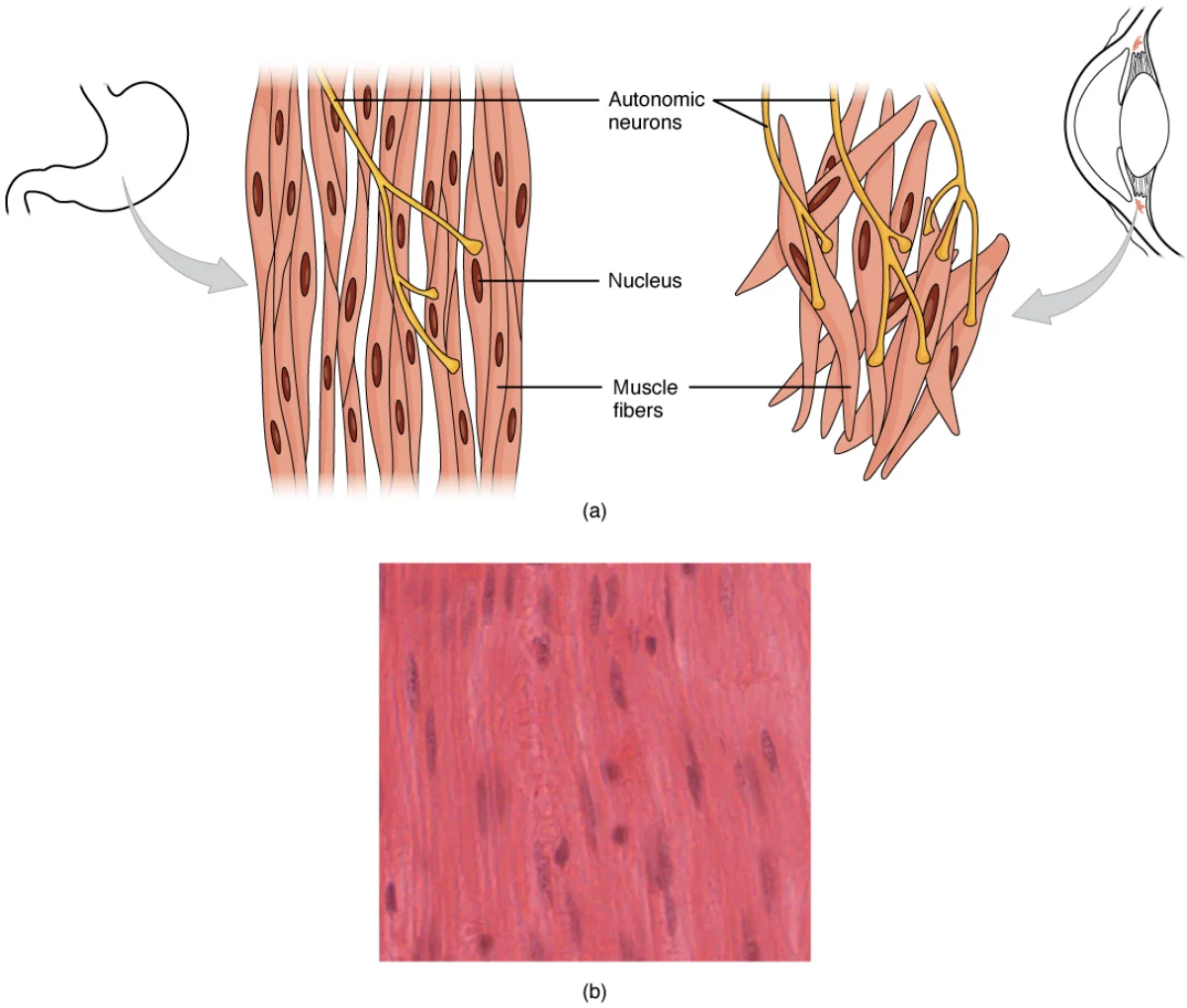
Smooth muscle gets its name from its lack of visible striations under the microscope. It does NOT have organized sarcomeres. Instead, contractile filaments are arranged in a criss-cross pattern anchored to structures called dense bodies (functionally similar to Z lines but scattered throughout the cell).
Smooth Muscle Contraction Mechanism
Smooth muscle uses a fundamentally different calcium-signaling pathway than skeletal muscle:
Ca²⁺ enters the cell from both the SR and the extracellular space
The Ca²⁺-calmodulin complex activates myosin light chain kinase (MLCK)
MLCK phosphorylates the myosin light chain, which activates the myosin ATPase
Phosphorylated myosin can now bind actin and perform cross-bridge cycling
Relaxation occurs when the myosin light chain is dephosphorylated
Two Types of Smooth Muscle
Feature
Single-Unit (Visceral)
Multi-Unit
Location
GI tract, uterus, ureter, bladder
Large airways, large arteries, iris, ciliary body, arrector pili
Gap junctions
Yes - cells connected electrically
No - each cell independently innervated
Contraction
Synchronized (cells contract as a sheet)
Independent (fine control)
Pacemaker activity
Yes (spontaneous depolarization)
No (needs nerve/hormone stimulation)
Stretch response
Contracts when stretched (myogenic response)
Minimal stretch response
Cardiac Muscle
Cardiac muscle shares features with both skeletal and smooth muscle. Like skeletal muscle, it has sarcomeres and striations. Like smooth muscle, it is involuntary and fatigue-resistant.
Unique Features of Cardiac Muscle
Intercalated discs are the defining structural feature of cardiac muscle. These are specialized junctions at the end-to-end connections between adjacent cardiac muscle cells. Each intercalated disc contains:
Gap junctions - allow ions to flow directly between cells, enabling the entire heart to contract as a synchronized unit (functional syncytium). When one cell depolarizes, the signal spreads to all connected cells.
Desmosomes - anchor cells together mechanically, preventing them from pulling apart during contraction
Smooth muscle cells are spindle-shaped with a single central nucleus and lack visible striations. Dense bodies (functionally analogous to Z lines) anchor the contractile filaments. Credit: OpenStax Anatomy and Physiology 2e, CC BY 4.0
Cardiac muscle uses a modified version of excitation-contraction coupling called calcium-induced calcium release (CICR):
Action potential propagates along the sarcolemma and into T-tubules
Calcium channels on T-tubules open and allow extracellular Ca²⁺ to enter the cell (unlike skeletal muscle, where the T-tubule acts as a mechanical sensor only)
This small influx of Ca²⁺ triggers calcium release channels on the SR to open
A much larger release of Ca²⁺ floods out of the SR (the “calcium-induced” part)
Ca²⁺ binds troponin C → cross-bridge cycling → contraction
The key difference from skeletal muscle: in cardiac muscle, extracellular Ca²⁺ is required. Block the calcium channels on the T-tubule, and the heart weakens its contraction.
Cardiac Muscle Cannot Tetanize
Skeletal muscle can sustain maximal contraction (tetanus) through rapid, repeated stimulation. Cardiac muscle CANNOT tetanize because it has a long refractory period that nearly spans the entire contraction. By the time the muscle is ready to respond to another stimulus, it has already relaxed. This is a critical safety mechanism - if the heart could tetanize, it would stop pumping blood and you would die.
In smooth muscle, what replaces troponin and tropomyosin as the calcium-sensitive regulatory system?
Click to reveal answer
Smooth muscle uses the calmodulin-MLCK system. Ca²⁺ binds to calmodulin (instead of troponin). The Ca²⁺-calmodulin complex activates myosin light chain kinase (MLCK), which phosphorylates the myosin regulatory light chain. This phosphorylation activates the myosin ATPase, allowing cross-bridge cycling. Unlike skeletal muscle (which regulates the thin filament), smooth muscle regulates the thick filament (myosin itself).
Why can't cardiac muscle undergo tetanus, and why is this physiologically important?
Click to reveal answer
Cardiac muscle has a long refractory period that lasts almost as long as the contraction itself. This means the cell cannot be re-stimulated until it has nearly finished relaxing, preventing summation of contractions. This is essential because tetanus in cardiac muscle would mean the heart stays contracted and stops pumping blood. The long refractory period ensures the heart contracts and relaxes rhythmically, maintaining blood flow.
The neuromuscular junction (NMJ) is where the nervous system meets the muscular system. It is the synapse between a motor neuron and a skeletal muscle fiber. Every voluntary movement begins here, and several major toxins, drugs, and diseases target this junction - making it a favorite MCAT topic.
Structure of the NMJ
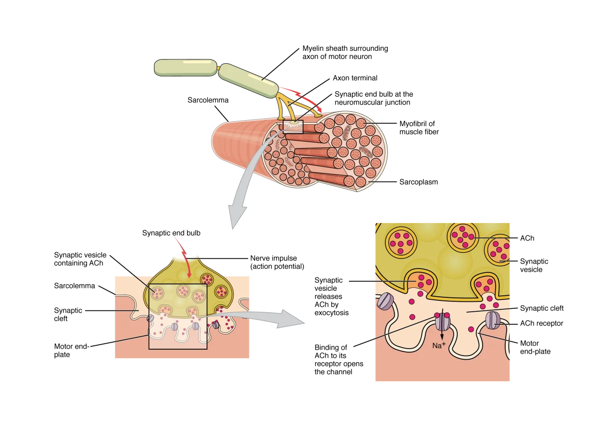
The neuromuscular junction. The motor neuron's axon terminal contains vesicles of ACh. When an action potential arrives, ACh is released into the synaptic cleft and binds nicotinic receptors on the motor end plate, triggering muscle fiber depolarization. Credit: OpenStax Anatomy and Physiology 2e, CC BY 4.0
The NMJ has three main components:
Axon terminal (synaptic bouton) - the end of the motor neuron axon, packed with synaptic vesicles containing acetylcholine (ACh). It also contains many mitochondria (for ATP production needed for vesicle packaging and recycling).
Synaptic cleft - the narrow gap (~50 nm) between the axon terminal and the muscle fiber. ACh diffuses across this gap. The enzyme acetylcholinesterase (AChE) is anchored in the synaptic cleft and rapidly breaks down ACh.
Motor end plate - the specialized region of the muscle fiber’s sarcolemma directly beneath the axon terminal. It is folded into junctional folds that increase surface area and concentrate nicotinic ACh receptors at the crests of the folds. Voltage-gated Na⁺ channels concentrate in the troughs.
Signal Transmission at the NMJ
Step 1: An action potential arrives at the axon terminal of the motor neuron.
Step 2: Voltage-gated Ca²⁺ channels open. Ca²⁺ flows into the axon terminal.
Step 3: Ca²⁺ triggers exocytosis of synaptic vesicles. ACh is released into the synaptic cleft. This calcium-dependent vesicle release follows the same principles as synaptic transmission at neuron-to-neuron synapses.
Step 4: ACh diffuses across the cleft and binds to nicotinic receptors on the motor end plate. These are ligand-gated ion channels that open when ACh binds, allowing Na⁺ to flow in (and some K⁺ out).
Step 5: Na⁺ influx depolarizes the motor end plate, producing an end plate potential (EPP). The EPP is always large enough to reach threshold, so every nerve impulse produces a muscle action potential. (There is no such thing as a “subthreshold” EPP under normal conditions.)
Step 6: The action potential propagates across the sarcolemma and into T-tubules, triggering E-C coupling and contraction.
Step 7: ACh is rapidly hydrolyzed by acetylcholinesterase (AChE) in the synaptic cleft:
ACh → choline + acetate. Choline is recycled back into the axon terminal for new ACh synthesis.
Drugs and Toxins That Target the NMJ
The NMJ is a target for many clinically important substances. The MCAT frequently presents passages about these:
Agent
Mechanism
Effect
Botulinum toxin (Botox)
Blocks ACh vesicle release
Paralysis (flaccid - muscle cannot contract)
Curare
Competitive antagonist at nicotinic receptors (blocks ACh binding)
A motor unit consists of one motor neuron and all the muscle fibers it innervates. The size of the motor unit determines the precision of control:
Small motor units (5-10 fibers per neuron) - found in muscles requiring fine control: fingers, eyes, tongue
Large motor units (hundreds of fibers per neuron) - found in muscles generating gross force: quadriceps, gastrocnemius
When the motor neuron fires, ALL fibers in that motor unit contract (all-or-none at the motor unit level). To increase force, the nervous system recruits additional motor units (recruitment). Motor units are recruited in order from smallest to largest (size principle).
A patient is exposed to an organophosphate nerve agent. What happens at the neuromuscular junction, and what symptoms would you expect?
Click to reveal answer
Organophosphates irreversibly inhibit acetylcholinesterase (AChE), the enzyme that breaks down ACh in the synaptic cleft. ACh accumulates, causing continuous stimulation of nicotinic receptors at the NMJ. This leads to sustained depolarization and muscle contraction that cannot stop. The key concept: if ACh is not removed from the cleft, the signal never turns off.
Why do muscles in the hand have small motor units while the quadriceps has large motor units?
Click to reveal answer
Motor unit size reflects the precision of control needed. The hand requires fine, precise movements (writing, typing, surgery), so each motor neuron controls only a few muscle fibers - this allows fine gradation of force. The quadriceps generates large, powerful movements (jumping, climbing) where fine control is less important, so each motor neuron controls hundreds of fibers, allowing efficient generation of large forces.
Your skin is the largest organ in your body, weighing about 4 kg (roughly 9 pounds) and covering approximately 2 square meters. It is far more than a passive wrapper - it is an active barrier, sensory organ, immune defense, vitamin D factory, and thermoregulation system all rolled into one.
The Three Layers of Skin
Layer
Type of Tissue
Key Features
Epidermis
Stratified squamous epithelium (keratinized)
Avascular, contains keratinocytes and melanocytes, outer protective layer
Not technically part of the skin; insulation, energy storage, cushioning
The Epidermis
The epidermis is the outermost layer. It is avascular (no blood supply) and composed of multiple layers of keratinocytes - cells that produce the tough protein keratin. As keratinocytes divide at the base and move upward, they gradually fill with keratin, flatten, and die. The outermost layer is composed entirely of dead, keratin-packed cells.
The two layers you need to know for the MCAT:
Stratum basale (basal layer) - the deepest layer. Contains a single row of stem cells that divide continuously to replenish the epidermis. Also contains melanocytes (produce the pigment melanin) and is attached to the basement membrane.
Stratum corneum - the outermost layer. Composed of 20-30 layers of dead, flattened, keratin-packed cells. This is the primary physical barrier and waterproof layer. These cells are continuously shed and replaced (the entire epidermis turns over roughly every 30 days).
Between these two layers, keratinocytes gradually fill with keratin, lose their organelles, and die as they move from the basal layer toward the surface.
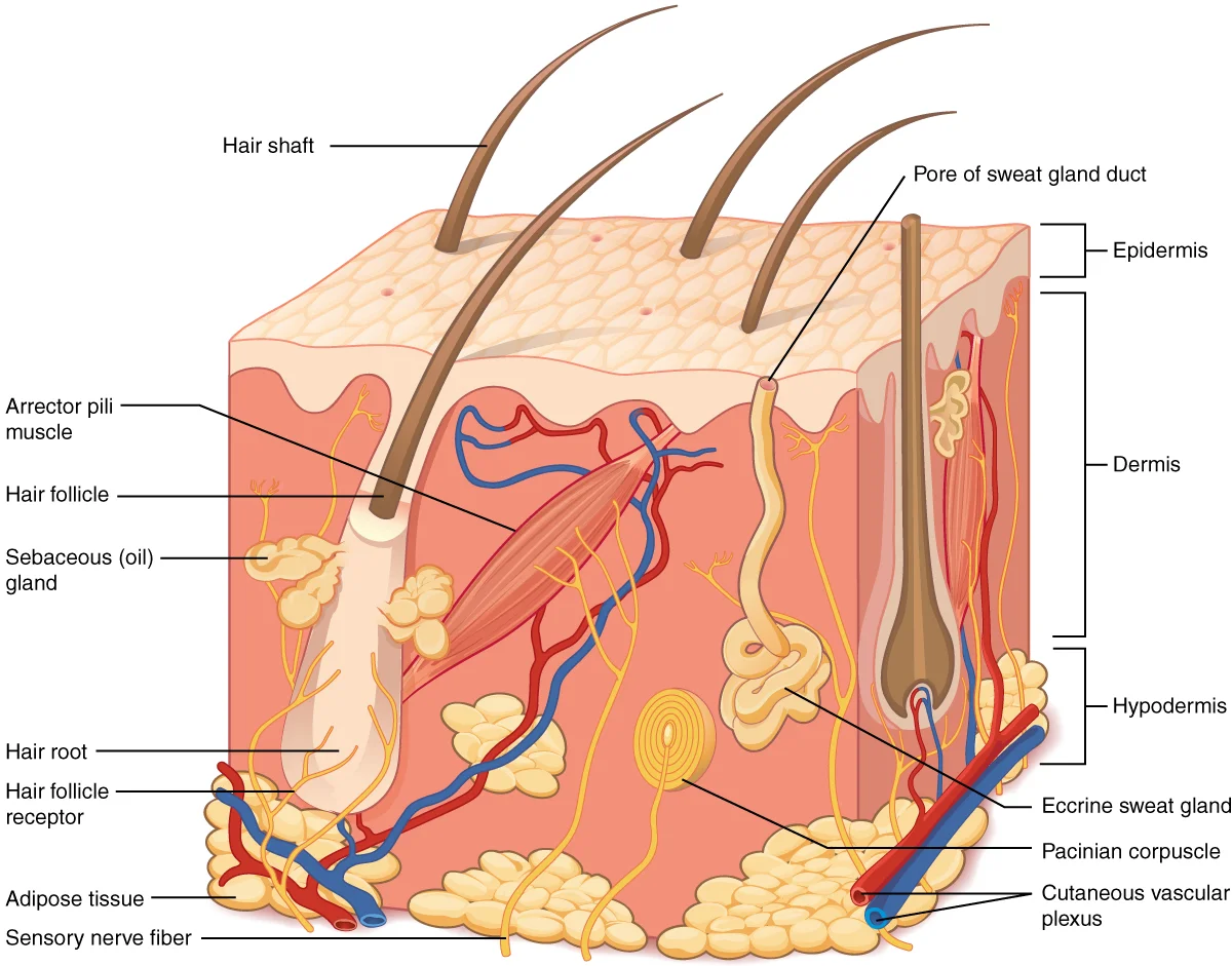
The layers of the skin. The epidermis sits on top of the dermis, which contains blood vessels, nerves, glands, and hair follicles. The hypodermis provides insulation and cushioning. Credit: OpenStax Anatomy and Physiology 2e, CC BY 4.0
Cells of the Epidermis
Cell Type
Function
Keratinocytes
Produce keratin; form the structural bulk of the epidermis (~90% of epidermal cells)
Melanocytes
Produce melanin (pigment that absorbs UV radiation); transfer melanin to surrounding keratinocytes
The Dermis
The dermis is the thick, structural layer beneath the epidermis. Unlike the epidermis, the dermis is vascularized (has blood vessels) and innervated (has nerves). It is made of dense irregular connective tissue containing collagen and elastin fibers, which give skin its strength and elasticity.
The dermis contains:
Blood vessels - supply nutrients to the epidermis (via diffusion) and play a major role in thermoregulation
Nerve endings and sensory receptors - detect touch, pressure, temperature, and pain
Hair follicles and arrector pili muscles (smooth muscle)
Sweat glands and sebaceous (oil) glands
Skin Appendages
Structure
Function
Notes
Hair follicles
Protection (head), sensation (hair movement triggers nerve endings)
Arrector pili muscles (smooth muscle) cause “goosebumps”
Sebaceous (oil) glands
Secrete sebum (oily substance that waterproofs and softens skin/hair)
Associated with hair follicles; holocrine secretion
Eccrine sweat glands
Thermoregulation via evaporative cooling
Found over entire body surface; secrete watery sweat directly onto skin surface via pores
Apocrine sweat glands
Produce thicker secretion (sweat + proteins/lipids)
Found in axillae and groin; empty into hair follicles; activated at puberty; bacterial breakdown of secretion causes body odor
Nails
Protect fingertips; assist with fine manipulation
Made of hard keratin; grow from the nail matrix
Thermoregulation
The skin is the body’s primary thermoregulatory organ. It uses several mechanisms:
When the body is too HOT:
Vasodilation of dermal blood vessels brings more warm blood near the surface, radiating heat outward
Eccrine sweat glands produce sweat; evaporation of water from the skin surface removes heat (evaporative cooling)
When the body is too COLD:
Vasoconstriction of dermal blood vessels reduces blood flow near the surface, conserving heat in the core
Arrector pili muscles contract (goosebumps) - minimally effective in humans but traps insulating air in furry animals
Shivering (controlled by skeletal muscle, not skin) generates heat through rapid, involuntary contractions
Vitamin D Synthesis
The skin initiates vitamin D synthesis. UV-B radiation converts a cholesterol precursor in the epidermis to vitamin D₃. This inactive form is then modified in the liver and again in the kidney to produce the active form, calcitriol. Calcitriol promotes calcium absorption in the intestines - connecting the skin directly to calcium homeostasis and bone health.
Which layer of the epidermis contains stem cells that continuously divide, and what specialized cells are also found there?
Click to reveal answer
The stratum basale (basal layer) is the deepest layer and contains keratinocyte stem cells that divide to replenish the epidermis. It also contains melanocytes, which produce melanin pigment. The stratum basale is the only layer with active cell division - all other epidermal cells are in various stages of differentiation and death as they move toward the surface.
How does the body increase heat loss through the skin when core temperature rises?
Click to reveal answer
Two primary mechanisms: (1) Vasodilation of dermal blood vessels increases blood flow near the skin surface, allowing more heat to radiate to the environment. (2) Eccrine sweat glands produce sweat, and the evaporation of water from the skin surface removes heat (evaporative cooling). Both are regulated by the hypothalamus and the autonomic nervous system.