
Sliding Filament Model
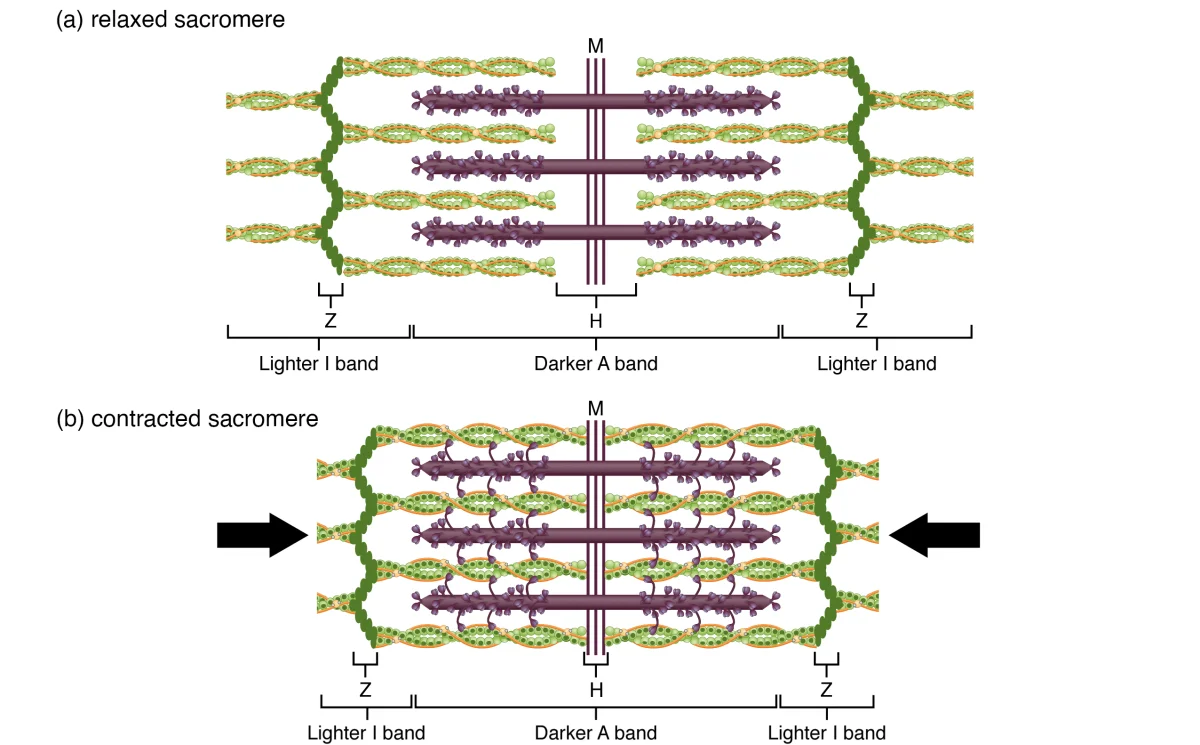
Here is the central question of muscle physiology: how does a muscle generate force? The answer is the sliding filament model - the most important concept in this entire chapter. The filaments themselves do not shorten. Instead, the thin filaments slide over the thick filaments, pulling the Z lines closer together and shortening the sarcomere. Multiply this across millions of sarcomeres contracting simultaneously, and you get the gross movement of the whole muscle.
Key Principles of the Sliding Filament Model
-
Filaments do NOT shorten. The actin and myosin filaments maintain their length throughout contraction. The sarcomere shortens because the filaments slide past each other.
-
The A band does not change width. The A band spans the full length of the thick filaments, which do not change length.
-
The I band and H zone shrink. As thin filaments slide inward over thick filaments, the regions containing only one type of filament get smaller.
-
Force is generated by cross-bridge cycling - the repeated attachment, pivoting, and detachment of myosin heads on actin filaments.
-
ATP is required for both contraction AND relaxation. ATP powers the myosin head reset and is required for myosin to release from actin.
Cross-Bridge Cycling - The Molecular Motor
Cross-bridge cycling is the step-by-step process by which myosin heads generate force on actin. Each cycle produces a small “power stroke” that pulls the thin filament toward the center of the sarcomere.
The Four Steps of Cross-Bridge Cycling
| Step | What Happens | Energy Source | State of Myosin Head |
|---|---|---|---|
| 1. Cross-bridge formation | Myosin head (energized, cocked position) binds to exposed binding site on actin | None (uses energy stored from previous step) | Bound to actin, high-energy conformation |
| 2. Power stroke | Myosin head pivots, pulling the thin filament toward the M line. ADP and Pi are released. | Stored elastic energy (from ATP hydrolysis in step 4) | Bound to actin, low-energy conformation |
| 3. Cross-bridge detachment | A new ATP molecule binds to the myosin head, causing it to release from actin | ATP binding (NOT hydrolysis) | Detached from actin, low-energy |
| 4. Myosin head reset (re-cocking) | Myosin’s ATPase hydrolyzes ATP → ADP + Pi. The energy is used to return the myosin head to its high-energy (cocked) position. | ATP hydrolysis | Detached from actin, high-energy (ready for next cycle) |
Then the cycle repeats from Step 1, as long as Ca²⁺ is present (keeping binding sites on actin exposed) and ATP is available.
The Role of ATP in Contraction
ATP plays TWO critical roles in muscle contraction:
-
Detachment - ATP binding to the myosin head breaks the cross-bridge (allows myosin to release from actin). Without ATP, myosin remains locked on actin.
-
Re-cocking - ATP hydrolysis (ATP → ADP + Pi) provides the energy to return the myosin head to its high-energy position, ready for the next power stroke.
Notice that ATP is NOT used during the power stroke itself. The power stroke is powered by the elastic energy that was stored in the myosin head during the previous hydrolysis step. The ATP is used to “reload the spring.”
The Role of Calcium in Contraction
Ca²⁺ is the ON switch for muscle contraction. Here is the sequence:
- At rest, tropomyosin covers the myosin-binding sites on actin. Cross-bridges cannot form.
- When Ca²⁺ is released from the sarcoplasmic reticulum, it binds to troponin C.
- Troponin changes shape, pulling tropomyosin away from the binding sites.
- Myosin-binding sites are now exposed, and cross-bridge cycling begins.
- When Ca²⁺ is pumped back into the SR (by the SERCA pump, which requires ATP), troponin releases Ca²⁺, tropomyosin slides back over the binding sites, and contraction stops.
The Length-Tension Relationship
The force a sarcomere generates depends on the degree of overlap between thick and thin filaments:
- Optimal overlap - maximum cross-bridges can form → maximum force
- Overstretched (too little overlap) - fewer cross-bridges can form → reduced force. At extreme stretch, no overlap → zero force.
- Over-compressed (too much overlap) - thin filaments from opposite sides interfere with each other, and thick filaments butt against Z lines → reduced force
This produces a bell-shaped length-tension curve. The MCAT may present this curve and ask you to explain why force changes at different sarcomere lengths.
The Force-Velocity Relationship
Force and velocity of contraction have an inverse relationship:
- Heavy load, slow contraction - when the load is large, each myosin head takes longer to pull the thin filament. Velocity drops toward zero as load approaches the muscle’s maximum force.
- Light load, fast contraction - when the load is small, thin filaments slide quickly past thick filaments. Velocity is highest with no load.
This is why you can lift a book fast but a heavy barbell slowly. The MCAT may show this hyperbolic curve and ask what happens when load changes.
The myosin head detaches from actin. ATP binding (not hydrolysis) causes a conformational change in the myosin head that breaks the cross-bridge. This is Step 3 of cross-bridge cycling. After detachment, the myosin ATPase hydrolyzes ATP → ADP + Pi, and the energy is used to re-cock the myosin head to its high-energy position (Step 4), preparing it for the next cycle.
Contraction requires ATP for two steps of cross-bridge cycling: (1) ATP binding detaches myosin from actin, and (2) ATP hydrolysis re-cocks the myosin head. Relaxation requires ATP because the SERCA pump (Ca²⁺-ATPase) on the sarcoplasmic reticulum uses ATP to actively pump Ca²⁺ back into the SR. Without this active transport, Ca²⁺ remains in the cytoplasm, troponin C stays bound to Ca²⁺, and the muscle stays contracted.