
Neuromuscular Junction
The neuromuscular junction (NMJ) is where the nervous system meets the muscular system. It is the synapse between a motor neuron and a skeletal muscle fiber. Every voluntary movement begins here, and several major toxins, drugs, and diseases target this junction - making it a favorite MCAT topic.
Structure of the NMJ
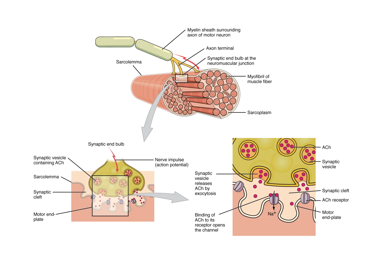
The NMJ has three main components:
-
Axon terminal (synaptic bouton) - the end of the motor neuron axon, packed with synaptic vesicles containing acetylcholine (ACh). It also contains many mitochondria (for ATP production needed for vesicle packaging and recycling).
-
Synaptic cleft - the narrow gap (~50 nm) between the axon terminal and the muscle fiber. ACh diffuses across this gap. The enzyme acetylcholinesterase (AChE) is anchored in the synaptic cleft and rapidly breaks down ACh.
-
Motor end plate - the specialized region of the muscle fiber’s sarcolemma directly beneath the axon terminal. It is folded into junctional folds that increase surface area and concentrate nicotinic ACh receptors at the crests of the folds. Voltage-gated Na⁺ channels concentrate in the troughs.
Signal Transmission at the NMJ
Step 1: An action potential arrives at the axon terminal of the motor neuron.
Step 2: Voltage-gated Ca²⁺ channels open. Ca²⁺ flows into the axon terminal.
Step 3: Ca²⁺ triggers exocytosis of synaptic vesicles. ACh is released into the synaptic cleft. This calcium-dependent vesicle release follows the same principles as synaptic transmission at neuron-to-neuron synapses.
Step 4: ACh diffuses across the cleft and binds to nicotinic receptors on the motor end plate. These are ligand-gated ion channels that open when ACh binds, allowing Na⁺ to flow in (and some K⁺ out).
Step 5: Na⁺ influx depolarizes the motor end plate, producing an end plate potential (EPP). The EPP is always large enough to reach threshold, so every nerve impulse produces a muscle action potential. (There is no such thing as a “subthreshold” EPP under normal conditions.)
Step 6: The action potential propagates across the sarcolemma and into T-tubules, triggering E-C coupling and contraction.
Step 7: ACh is rapidly hydrolyzed by acetylcholinesterase (AChE) in the synaptic cleft:
ACh → choline + acetate. Choline is recycled back into the axon terminal for new ACh synthesis.
Drugs and Toxins That Target the NMJ
The NMJ is a target for many clinically important substances. The MCAT frequently presents passages about these:
| Agent | Mechanism | Effect |
|---|---|---|
| Botulinum toxin (Botox) | Blocks ACh vesicle release | Paralysis (flaccid - muscle cannot contract) |
| Curare | Competitive antagonist at nicotinic receptors (blocks ACh binding) | Paralysis (flaccid) |
| Nerve agents (organophosphates) | Irreversibly inhibit AChE (ACh accumulates) | Overstimulation → sustained contraction → paralysis |
| AChE inhibitors (e.g., neostigmine) | Reversibly inhibit AChE (increases ACh in cleft) | Used to treat myasthenia gravis |
The Motor Unit
A motor unit consists of one motor neuron and all the muscle fibers it innervates. The size of the motor unit determines the precision of control:
- Small motor units (5-10 fibers per neuron) - found in muscles requiring fine control: fingers, eyes, tongue
- Large motor units (hundreds of fibers per neuron) - found in muscles generating gross force: quadriceps, gastrocnemius
When the motor neuron fires, ALL fibers in that motor unit contract (all-or-none at the motor unit level). To increase force, the nervous system recruits additional motor units (recruitment). Motor units are recruited in order from smallest to largest (size principle).