
Excitation-Contraction Coupling
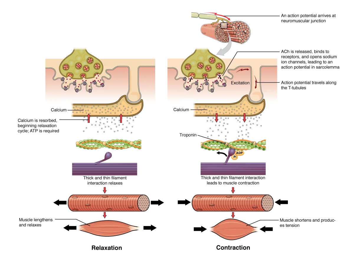
You now know the molecular machinery of contraction (cross-bridge cycling) and its on-switch (calcium). But how does a decision in your brain translate into calcium flooding a muscle cell? That is excitation-contraction (E-C) coupling - the chain of events that converts an electrical signal (action potential) into a mechanical response (muscle contraction).
The Complete E-C Coupling Pathway
Follow each step carefully - this sequence is one of the most commonly tested pathways on the MCAT:
Step 1: Action potential arrives at the neuromuscular junction
A motor neuron releases acetylcholine (ACh) into the synaptic cleft. ACh binds to nicotinic receptors on the sarcolemma (muscle cell membrane), opening Na⁺ channels and depolarizing the muscle fiber.
Step 2: Action potential propagates along the sarcolemma
The depolarization triggers a full action potential that spreads across the entire surface of the muscle fiber.
Step 3: Action potential dives into T-tubules
The action potential travels down the T-tubules (transverse tubules) - deep invaginations of the sarcolemma that penetrate into the interior of the fiber. This ensures the signal reaches every sarcomere simultaneously, even those deep inside the cell.
Step 4: Voltage sensor activates calcium release
The T-tubule membrane contains voltage-sensitive proteins called dihydropyridine (DHP) receptors (L-type calcium channels). When the action potential depolarizes the T-tubule, DHP receptors change conformation and physically interact with ryanodine receptors (RyR) on the adjacent sarcoplasmic reticulum membrane.
Step 5: SR releases calcium
Activated ryanodine receptors open, and Ca²⁺ floods out of the SR into the sarcoplasm. Ca²⁺ concentration in the sarcoplasm jumps from ~ M to ~ M (a 100-fold increase).
Step 6: Calcium triggers contraction
Ca²⁺ binds to troponin C on the thin filaments → tropomyosin shifts → myosin-binding sites on actin are exposed → cross-bridge cycling begins → the sarcomere shortens → the muscle contracts.
Step 7: Relaxation
When the nerve signal stops, ACh is broken down by acetylcholinesterase (AChE), and the action potential ceases. The SERCA pump (SR Ca²⁺-ATPase) actively pumps Ca²⁺ back into the sarcoplasmic reticulum. As cytoplasmic Ca²⁺ drops, Ca²⁺ dissociates from troponin C, tropomyosin slides back over the binding sites, and cross-bridge cycling stops. The muscle relaxes.
The Triad
The triad is the structural unit that makes E-C coupling work. It consists of:
- One T-tubule (carrying the electrical signal)
- Two terminal cisternae of the SR (calcium reservoirs flanking the T-tubule)
This arrangement ensures the voltage sensor (DHP receptor on the T-tubule) is physically adjacent to the calcium release channel (ryanodine receptor on the SR). The signal does not have to diffuse - it is transmitted by direct mechanical coupling.
Skeletal vs. Cardiac E-C Coupling
There is a critical difference between the two muscle types:
-
In skeletal muscle, the voltage sensor on the T-tubule is physically connected to the calcium release channel on the SR. The action potential triggers calcium release through direct mechanical coupling - no extracellular calcium needs to enter the cell.
-
In cardiac muscle, the T-tubule voltage sensor actually lets a small amount of extracellular Ca²⁺ into the cell. This small Ca²⁺ influx then triggers a much larger release of Ca²⁺ from the SR. This is called calcium-induced calcium release (CICR).
Summary: The Signal Chain
Here is the entire pathway condensed into one chain:
Motor neuron → ACh release → Nicotinic receptor on sarcolemma → Na⁺ influx → Action potential → T-tubule → DHP receptor → Ryanodine receptor → Ca²⁺ released from SR → Ca²⁺ binds troponin C → Tropomyosin moves → Cross-bridge cycling → CONTRACTION
And for relaxation:
ACh degraded by AChE → No more action potential → SERCA pump returns Ca²⁺ to SR → Troponin releases Ca²⁺ → Tropomyosin covers binding sites → RELAXATION
Muscle Twitch, Summation, and Tetanus
When a single action potential causes a single, brief contraction, that is a muscle twitch. But in real life, muscles rarely twitch once. Instead, motor neurons fire repeatedly:
-
Temporal summation (wave summation) - if a second stimulus arrives before the muscle fully relaxes from the first, the second contraction adds to the first, producing greater force. The muscle does not have time to fully relax between stimuli.
-
Incomplete tetanus - rapid, repeated stimulation where the muscle partially relaxes between contractions. Force is higher than a single twitch but fluctuates.
-
Complete tetanus - stimulation is so rapid that the muscle has no time to relax at all. The individual twitches fuse into one smooth, sustained contraction at maximum force. This is normal voluntary muscle contraction.
Motor unit recruitment also increases force. A motor unit is one motor neuron plus all the muscle fibers it innervates. To generate more force, the nervous system recruits additional motor units. Small motor units (few fibers) are recruited first for fine control; large motor units (many fibers) are added for powerful movements.